3OIX

Crystal structure of the putative dihydroorotate dehydrogenase from Streptococcus mutans


Experimental Data Snapshot

  • Method: X-RAY DIFFRACTION
  • Resolution: 2.40 Å
  • R-Value Free: 0.248 
  • R-Value Work: 0.202 
  • R-Value Observed: 0.204 

wwPDB Validation   3D Report Full Report


Ligand Structure Quality Assessment 


This is version 1.3 of the entry. See complete history


Literature

Structure of the putative dihydroorotate dehydrogenase from Streptococcus mutans

Liu, Y.Gao, Z.-Q.Liu, C.-P.Xu, J.-H.Li, L.-F.Ji, C.-N.Su, X.-D.Dong, Y.-H.

(2011) Acta Crystallogr Sect F Struct Biol Cryst Commun 67: 182-187

  • DOI: https://doi.org/10.1107/S1744309110048414
  • Primary Citation of Related Structures:  
    3OIX

  • PubMed Abstract: 

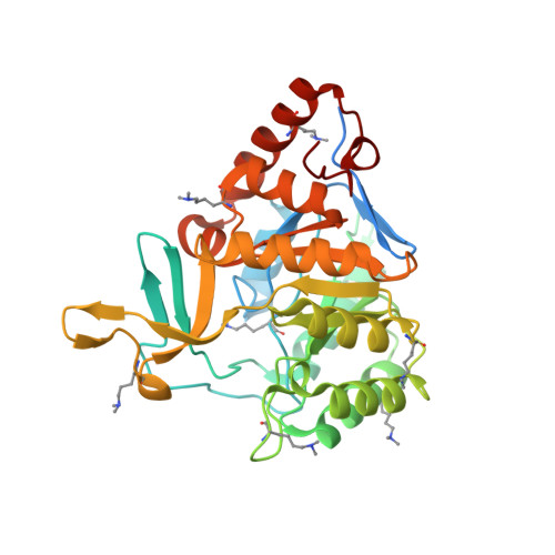
    Streptococcus mutans is one of the pathogenic species involved in dental caries, especially in the initiation and development stages. Here, the crystal structure of SMU.595, a putative dihydroorotate dehydrogenase (DHOD) from S. mutans, is reported at 2.4 Å resolution. DHOD is a flavin mononucleotide-containing enzyme which catalyzes the oxidation of L-dihydroorotate to orotate, which is the fourth step and the only redox reaction in the de novo biosynthesis of pyrimidine nucleotides. The reductive lysine-methylation procedure was applied in order to improve the diffraction qualities of the crystals. Analysis of the S. mutans DHOD crystal structure shows that this enzyme is a class 1A DHOD and also suggests potential sites that could be exploited for the design of highly specific inhibitors using the structure-based chemotherapeutic design technique.


  • Organizational Affiliation

    Department of Genetics, School of Life Science, Fudan University, Shanghai, People's Republic of China.


Macromolecules
Find similar proteins by:  (by identity cutoff)  |  3D Structure
Entity ID: 1
MoleculeChains Sequence LengthOrganismDetailsImage
Putative dihydroorotate dehydrogenase; dihydroorotate oxidase
A, B, C, D
345Streptococcus mutansMutation(s): 0 
Gene Names: smu.595
EC: 1.3.3.1 (PDB Primary Data), 1.3.5.2 (PDB Primary Data)
UniProt
Find proteins for Q8DVA1 (Streptococcus mutans serotype c (strain ATCC 700610 / UA159))
Explore Q8DVA1 
Go to UniProtKB:  Q8DVA1
Entity Groups  
Sequence Clusters30% Identity50% Identity70% Identity90% Identity95% Identity100% Identity
UniProt GroupQ8DVA1
Sequence Annotations
Expand
  • Reference Sequence
Experimental Data & Validation

Experimental Data

  • Method: X-RAY DIFFRACTION
  • Resolution: 2.40 Å
  • R-Value Free: 0.248 
  • R-Value Work: 0.202 
  • R-Value Observed: 0.204 
  • Space Group: C 2 2 21
Unit Cell:
Length ( Å )Angle ( ˚ )
a = 103.126α = 90
b = 158.276β = 90
c = 193.351γ = 90
Software Package:
Software NamePurpose
MAR345data collection
PHASERphasing
PHENIXrefinement
HKL-2000data reduction
HKL-2000data scaling

Structure Validation

View Full Validation Report


Ligand Structure Quality Assessment 


Entry History 

Deposition Data

Revision History  (Full details and data files)

  • Version 1.0: 2010-09-08
    Type: Initial release
  • Version 1.1: 2011-07-13
    Changes: Version format compliance
  • Version 1.2: 2017-11-08
    Changes: Refinement description
  • Version 1.3: 2023-11-01
    Changes: Data collection, Database references, Derived calculations, Refinement description