3OB4

MBP-fusion protein of the major peanut allergen Ara h 2


Experimental Data Snapshot

  • Method: X-RAY DIFFRACTION
  • Resolution: 2.71 Å
  • R-Value Free: 0.256 
  • R-Value Work: 0.183 
  • R-Value Observed: 0.187 

wwPDB Validation   3D Report Full Report


This is version 2.2 of the entry. See complete history


Literature

Ara h 2: crystal structure and IgE binding distinguish two subpopulations of peanut allergic patients by epitope diversity.

Mueller, G.A.Gosavi, R.A.Pomes, A.Wunschmann, S.Moon, A.F.London, R.E.Pedersen, L.C.

(2011) Allergy 66: 878-885

  • DOI: https://doi.org/10.1111/j.1398-9995.2010.02532.x
  • Primary Citation of Related Structures:  
    3OB4

  • PubMed Abstract: 

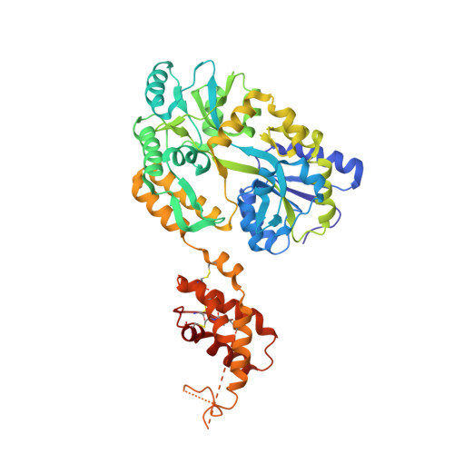
    Peanut allergy affects 1% of the population and causes the most fatal food-related anaphylactic reactions. The protein Ara h 2 is the most potent peanut allergen recognized by 80-90% of peanut allergic patients. The crystal structure of the major peanut allergen Ara h 2 was determined for the first time at 2.7 Å resolution using a customized maltose-binding protein (MBP)-fusion system. IgE antibody binding to the MBP fusion construct vs the natural allergen was compared by ELISA using sera from peanut allergic patients. The structure of Ara h 2 is a five-helix bundle held together by four disulfide bonds and related to the prolamin protein superfamily. The fold is most similar to other amylase and trypsin inhibitors. The MBP--Ara h 2 fusion construct was positively recognized by IgE from 76% of allergic patients (25/33). Two populations of patients could be identified. Subpopulation 1 (n = 14) showed an excellent correlation of IgE antibody binding to natural vs recombinant Ara h 2. Subpopulation 2 (n = 15) showed significantly reduced IgE binding to the MBP fusion protein. Interestingly, about 20% of the IgE binding in subpopulation 2 could be recovered by increasing the distance between MBP and Ara h 2 in a second construct. The reduced IgE binding to the MBP--Ara h 2 of subpopulation 2 indicates that the MBP molecule protects an immunodominant epitope region near the first helix of Ara h 2. Residues involved in the epitope(s) are suggested by the crystal structure. The MBP--Ara h 2 fusion constructs will be useful to further elucidate the relevance of certain epitopes to peanut allergy.


  • Organizational Affiliation

    Laboratory of Structural Biology, National Institute of Environmental Health Sciences, Research Triangle Park, NC, USA. mueller3@niehs.nih.gov


Macromolecules
Find similar proteins by:  (by identity cutoff)  |  3D Structure
Entity ID: 1
MoleculeChains Sequence LengthOrganismDetailsImage
Maltose ABC transporter periplasmic protein,Arah 2500Escherichia coliArachis hypogaea
This entity is chimeric
Mutation(s): 7 
Gene Names: HMPREF9530_03068Ara h 2
UniProt
Find proteins for P0AEX9 (Escherichia coli (strain K12))
Explore P0AEX9 
Go to UniProtKB:  P0AEX9
Find proteins for A5Z1Q8 (Arachis duranensis)
Explore A5Z1Q8 
Go to UniProtKB:  A5Z1Q8
Entity Groups  
Sequence Clusters30% Identity50% Identity70% Identity90% Identity95% Identity100% Identity
UniProt GroupsA5Z1Q8P0AEX9
Sequence Annotations
Expand
  • Reference Sequence
Oligosaccharides

Help

Entity ID: 2
MoleculeChains Length2D Diagram Glycosylation3D Interactions
alpha-D-glucopyranose-(1-4)-alpha-D-glucopyranose-(1-4)-alpha-D-glucopyranose
B
3N/A
Glycosylation Resources
GlyTouCan:  G96370VA
GlyCosmos:  G96370VA
GlyGen:  G96370VA
Biologically Interesting Molecules (External Reference) 1 Unique
Experimental Data & Validation

Experimental Data

  • Method: X-RAY DIFFRACTION
  • Resolution: 2.71 Å
  • R-Value Free: 0.256 
  • R-Value Work: 0.183 
  • R-Value Observed: 0.187 
  • Space Group: C 1 2 1
Unit Cell:
Length ( Å )Angle ( ˚ )
a = 68.847α = 90
b = 87.381β = 103.93
c = 113.137γ = 90
Software Package:
Software NamePurpose
MAR345data collection
MOLREPphasing
PHENIXrefinement
HKL-2000data reduction
HKL-2000data scaling

Structure Validation

View Full Validation Report


Entry History 

Deposition Data

Revision History  (Full details and data files)

  • Version 1.0: 2011-02-02
    Type: Initial release
  • Version 1.1: 2011-07-13
    Changes: Version format compliance
  • Version 1.2: 2017-08-09
    Changes: Refinement description, Source and taxonomy
  • Version 1.3: 2017-11-08
    Changes: Refinement description
  • Version 1.4: 2020-02-26
    Changes: Database references
  • Version 2.0: 2020-07-29
    Type: Remediation
    Reason: Carbohydrate remediation
    Changes: Atomic model, Data collection, Derived calculations, Non-polymer description, Structure summary
  • Version 2.1: 2021-02-17
    Changes: Database references, Source and taxonomy, Structure summary
  • Version 2.2: 2023-09-06
    Changes: Data collection, Database references, Refinement description