3O7B

Crystal structure of Archaeoglobus Fulgidus Nep1 bound to S-adenosylhomocysteine


Experimental Data Snapshot

  • Method: X-RAY DIFFRACTION
  • Resolution: 1.45 Å
  • R-Value Free: 0.211 
  • R-Value Work: 0.198 
  • R-Value Observed: 0.199 

wwPDB Validation   3D Report Full Report


Ligand Structure Quality Assessment 


This is version 1.2 of the entry. See complete history


Literature

Structural insight into the functional mechanism of Nep1/Emg1 N1-specific pseudouridine methyltransferase in ribosome biogenesis.

Thomas, S.R.Keller, C.A.Szyk, A.Cannon, J.R.Laronde-Leblanc, N.A.

(2011) Nucleic Acids Res 39: 2445-2457

  • DOI: https://doi.org/10.1093/nar/gkq1131
  • Primary Citation of Related Structures:  
    3O7B, 3OII, 3OIJ, 3OIN

  • PubMed Abstract: 

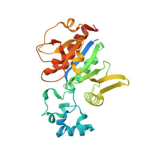
    Nucleolar Essential Protein 1 (Nep1) is required for small subunit (SSU) ribosomal RNA (rRNA) maturation and is mutated in Bowen-Conradi Syndrome. Although yeast (Saccharomyces cerevisiae) Nep1 interacts with a consensus sequence found in three regions of SSU rRNA, the molecular details of the interaction are unknown. Nep1 is a SPOUT RNA methyltransferase, and can catalyze methylation at the N1 of pseudouridine. Nep1 is also involved in assembly of Rps19, an SSU ribosomal protein. Mutations in Nep1 that result in decreased methyl donor binding do not result in lethality, suggesting that enzymatic activity may not be required for function, and RNA binding may play a more important role. To study these interactions, the crystal structures of the scNep1 dimer and its complexes with RNA were determined. The results demonstrate that Nep1 recognizes its RNA site via base-specific interactions and stabilizes a stem-loop in the bound RNA. Furthermore, the RNA structure observed contradicts the predicted structures of the Nep1-binding sites within mature rRNA, suggesting that the Nep1 changes rRNA structure upon binding. Finally, a uridine base is bound in the active site of Nep1, positioned for a methyltransfer at the C5 position, supporting its role as an N1-specific pseudouridine methyltransferase.


  • Organizational Affiliation

    Department of Chemistry and Biochemistry, Center for Biomolecular Structure and Organization, University of Maryland, College Park, MD 20742, USA.


Macromolecules
Find similar proteins by:  (by identity cutoff)  |  3D Structure
Entity ID: 1
MoleculeChains Sequence LengthOrganismDetailsImage
Ribosome biogenesis Nep1 RNA methyltransferase244Archaeoglobus fulgidusMutation(s): 0 
Gene Names: AF_0734
UniProt
Find proteins for O29524 (Archaeoglobus fulgidus (strain ATCC 49558 / DSM 4304 / JCM 9628 / NBRC 100126 / VC-16))
Explore O29524 
Go to UniProtKB:  O29524
Entity Groups  
Sequence Clusters30% Identity50% Identity70% Identity90% Identity95% Identity100% Identity
UniProt GroupO29524
Sequence Annotations
Expand
  • Reference Sequence
Small Molecules
Ligands 2 Unique
IDChains Name / Formula / InChI Key2D Diagram3D Interactions
SAH
Query on SAH

Download Ideal Coordinates CCD File 
C [auth A]S-ADENOSYL-L-HOMOCYSTEINE
C14 H20 N6 O5 S
ZJUKTBDSGOFHSH-WFMPWKQPSA-N
TYR
Query on TYR

Download Ideal Coordinates CCD File 
B [auth A]TYROSINE
C9 H11 N O3
OUYCCCASQSFEME-QMMMGPOBSA-N
Experimental Data & Validation

Experimental Data

  • Method: X-RAY DIFFRACTION
  • Resolution: 1.45 Å
  • R-Value Free: 0.211 
  • R-Value Work: 0.198 
  • R-Value Observed: 0.199 
  • Space Group: P 31 2 1
Unit Cell:
Length ( Å )Angle ( ˚ )
a = 70.083α = 90
b = 70.083β = 90
c = 95.19γ = 120
Software Package:
Software NamePurpose
DENZOdata reduction
SCALEPACKdata scaling
REFMACrefinement
PDB_EXTRACTdata extraction
SHARPphasing

Structure Validation

View Full Validation Report


Ligand Structure Quality Assessment 


Entry History 

Deposition Data

Revision History  (Full details and data files)

  • Version 1.0: 2011-01-19
    Type: Initial release
  • Version 1.1: 2011-07-13
    Changes: Version format compliance
  • Version 1.2: 2017-11-08
    Changes: Advisory, Refinement description