


Crystal structure of CAD domain of the Plasmodium Vivax CDPK, PVX_11610
Wernimont, A.K., Hutchinson, A., Sullivan, H., Weadge, J., Bochkarev, A., Arrowsmith, C.H., Edwards, A.M., Bountra, C., Weigelt, J., Hui, R., Amani, M.To be published.
Experimental Data Snapshot
wwPDB Validation 3D Report Full Report

Entity ID: 1 | |||||
|---|---|---|---|---|---|
| Molecule | Chains | Sequence Length | Organism | Details | Image |
| Calcium-dependent protein kinase 3 | 196 | Plasmodium vivax | Mutation(s): 0 Gene Names: PVX_119610 | 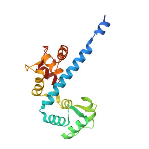 | |
UniProt | |||||
Find proteins for A5KBB4 (Plasmodium vivax (strain Salvador I)) Explore A5KBB4 Go to UniProtKB: A5KBB4 | |||||
Entity Groups | |||||
| Sequence Clusters | 30% Identity50% Identity70% Identity90% Identity95% Identity100% Identity | ||||
| UniProt Group | A5KBB4 | ||||
Sequence AnnotationsExpand | |||||
| |||||
| Ligands 3 Unique | |||||
|---|---|---|---|---|---|
| ID | Chains | Name / Formula / InChI Key | 2D Diagram | 3D Interactions | |
| PO4 Query on PO4 | E [auth A] | PHOSPHATE ION O4 P NBIIXXVUZAFLBC-UHFFFAOYSA-K |  | ||
| GOL Query on GOL | D [auth A] | GLYCEROL C3 H8 O3 PEDCQBHIVMGVHV-UHFFFAOYSA-N |  | ||
| CA Query on CA | B [auth A], C [auth A] | CALCIUM ION Ca BHPQYMZQTOCNFJ-UHFFFAOYSA-N |  | ||
| Length ( Å ) | Angle ( ˚ ) |
|---|---|
| a = 52.106 | α = 90 |
| b = 84.858 | β = 90 |
| c = 85.757 | γ = 90 |
| Software Name | Purpose |
|---|---|
| DENZO | data reduction |
| SCALEPACK | data scaling |
| REFMAC | refinement |
| PDB_EXTRACT | data extraction |
| SBC-Collect | data collection |
| PHASER | phasing |
RCSB PDB (citation) is hosted by
RCSB PDB is a member of the

