3O1A

Structure of OxyE (CYP165D3), a Cytochrome P450 Involved in Teicoplanin Biosynthesis


Experimental Data Snapshot

  • Method: X-RAY DIFFRACTION
  • Resolution: 2.50 Å
  • R-Value Free: 0.274 
  • R-Value Work: 0.220 
  • R-Value Observed: 0.223 

wwPDB Validation   3D Report Full Report


Ligand Structure Quality Assessment 


This is version 1.2 of the entry. See complete history


Literature

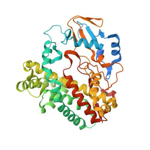
Structural characterization of CYP165D3, a cytochrome P450 involved in phenolic coupling in teicoplanin biosynthesis.

Cryle, M.J.Staaden, J.Schlichting, I.

(2011) Arch Biochem Biophys 507: 163-173

  • DOI: https://doi.org/10.1016/j.abb.2010.10.017
  • Primary Citation of Related Structures:  
    3O1A

  • PubMed Abstract: 

    Teicoplanin is a glycopeptide antibiotic with activity against Gram-positive bacteria and remains one of the last lines of clinical defense against certain bacterial infections. We have cloned, expressed, and purified the cytochrome P450 OxyE (CYP165D3) from the teicoplanin biosynthetic gene cluster of Actinoplanes teichomyceticus, which is responsible for the phenolic coupling of the aromatic side chains of the first and third peptide residues in the teicoplanin peptide. The crystal structure of OxyE has been determined to 2.5Å resolution, revealing the probable binding surface for the carrier protein substrate and an extension of the active site into a pocket located above the β-1 sheet. The binding of potential substrates to OxyE shows that peptidyl carrier protein-bound linear peptides bind to OxyE, albeit with low affinity in the absence of a phenolic cross-link that should normally be installed by another Oxy protein in the teicoplanin biosynthetic pathway. This result indicates that the carrier protein alone is not sufficient for tight substrate binding to OxyE and that the Oxy proteins sense the structure of the bound peptide in addition to the presence of the carrier protein, a feature distinct from other carrier protein/P450 systems.


  • Organizational Affiliation

    Department of Biomolecular Mechanisms, Max-Planck Institute for Medical Research, Jahnstrasse 29, Heidelberg, Germany. Max.Cryle@mpimf-heidelberg.mpg.de


Macromolecules
Find similar proteins by:  (by identity cutoff)  |  3D Structure
Entity ID: 1
MoleculeChains Sequence LengthOrganismDetailsImage
Oxy protein417Actinoplanes teichomyceticusMutation(s): 0 
Gene Names: tcp18tcp19
EC: 1.14.14.1
UniProt
Find proteins for Q6ZZI7 (Actinoplanes teichomyceticus)
Explore Q6ZZI7 
Go to UniProtKB:  Q6ZZI7
Entity Groups  
Sequence Clusters30% Identity50% Identity70% Identity90% Identity95% Identity100% Identity
UniProt GroupQ6ZZI7
Sequence Annotations
Expand
  • Reference Sequence
Small Molecules
Ligands 1 Unique
IDChains Name / Formula / InChI Key2D Diagram3D Interactions
HEM
Query on HEM

Download Ideal Coordinates CCD File 
B [auth A]PROTOPORPHYRIN IX CONTAINING FE
C34 H32 Fe N4 O4
KABFMIBPWCXCRK-RGGAHWMASA-L
Experimental Data & Validation

Experimental Data

  • Method: X-RAY DIFFRACTION
  • Resolution: 2.50 Å
  • R-Value Free: 0.274 
  • R-Value Work: 0.220 
  • R-Value Observed: 0.223 
  • Space Group: P 31
Unit Cell:
Length ( Å )Angle ( ˚ )
a = 75.68α = 90
b = 75.68β = 90
c = 74.72γ = 120
Software Package:
Software NamePurpose
XDSdata scaling
PHASERphasing
REFMACrefinement
XDSdata reduction
XSCALEdata scaling

Structure Validation

View Full Validation Report


Ligand Structure Quality Assessment 


Entry History 

Deposition Data

Revision History  (Full details and data files)

  • Version 1.0: 2010-11-03
    Type: Initial release
  • Version 1.1: 2011-07-13
    Changes: Version format compliance
  • Version 1.2: 2023-09-06
    Changes: Data collection, Database references, Derived calculations, Refinement description