


Crystal structure of anti-emmprin antibody 5F6 FAB
Teplyakov, A., Obmolova, G., Malia, T., Gilliland, G.L.To be published.
Experimental Data Snapshot
wwPDB Validation 3D Report Full Report

Entity ID: 1 | |||||
|---|---|---|---|---|---|
| Molecule | Chains | Sequence Length | Organism | Details | Image |
| 5F6 LIGHT CHAIN | A [auth L] | 214 | Homo sapiens, Mus musculus This entity is chimeric | Mutation(s): 0 | 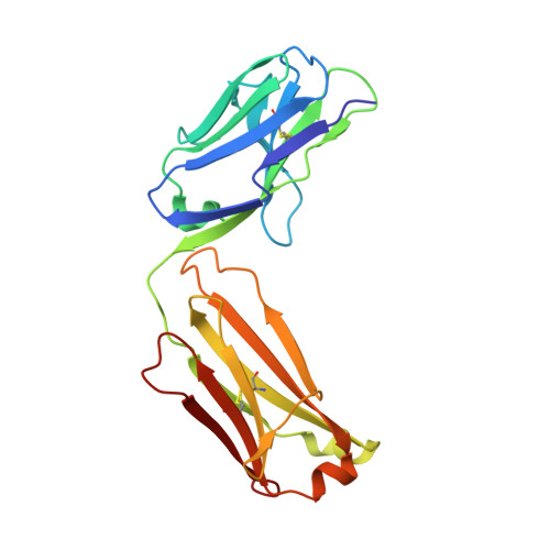 |
Entity Groups | |||||
| Sequence Clusters | 30% Identity50% Identity70% Identity90% Identity95% Identity100% Identity | ||||
Sequence AnnotationsExpand | |||||
| |||||
Entity ID: 2 | |||||
|---|---|---|---|---|---|
| Molecule | Chains | Sequence Length | Organism | Details | Image |
| 5F6 HEAVY CHAIN | B [auth H] | 226 | Homo sapiens, Mus musculus This entity is chimeric | Mutation(s): 0 | 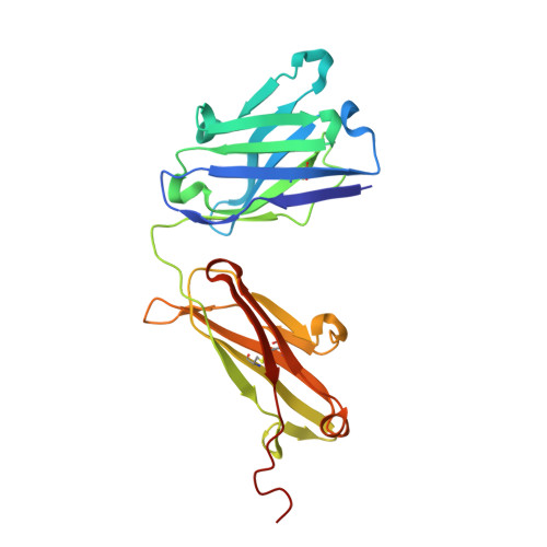 |
Entity Groups | |||||
| Sequence Clusters | 30% Identity50% Identity70% Identity90% Identity95% Identity100% Identity | ||||
Sequence AnnotationsExpand | |||||
| |||||
| Ligands 2 Unique | |||||
|---|---|---|---|---|---|
| ID | Chains | Name / Formula / InChI Key | 2D Diagram | 3D Interactions | |
| GOL Query on GOL | C [auth L] | GLYCEROL C3 H8 O3 PEDCQBHIVMGVHV-UHFFFAOYSA-N |  | ||
| CO Query on CO | D [auth H] | COBALT (II) ION Co XLJKHNWPARRRJB-UHFFFAOYSA-N |  | ||
| Length ( Å ) | Angle ( ˚ ) |
|---|---|
| a = 96.53 | α = 90 |
| b = 129.74 | β = 109.52 |
| c = 42.39 | γ = 90 |
| Software Name | Purpose |
|---|---|
| CrystalClear | data collection |
| PHASER | phasing |
| REFMAC | refinement |
| d*TREK | data reduction |
| d*TREK | data scaling |
RCSB PDB (citation) is hosted by
RCSB PDB is a member of the

