3NV5

Crystal Structure of Cytochrome P450 CYP101D2


Experimental Data Snapshot

  • Method: X-RAY DIFFRACTION
  • Resolution: 2.41 Å
  • R-Value Free: 0.260 
  • R-Value Work: 0.196 
  • R-Value Observed: 0.199 

wwPDB Validation   3D Report Full Report


Ligand Structure Quality Assessment 


This is version 1.3 of the entry. See complete history


Literature

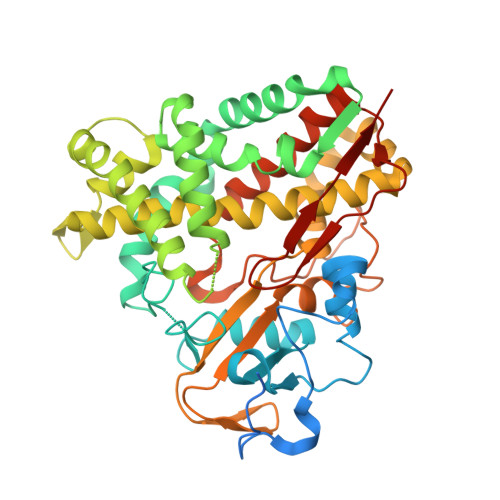
The structure of CYP101D2 unveils a potential path for substrate entry into the active site

Yang, W.Bell, S.G.Wang, H.Zhou, W.Bartlam, M.Wong, L.L.Rao, Z.

(2011) Biochem J 433: 85-93

  • DOI: https://doi.org/10.1042/BJ20101017
  • Primary Citation of Related Structures:  
    3NV5, 3NV6

  • PubMed Abstract: 

    The cytochrome P450 CYP101D2 from Novosphingobium aromaticivorans DSM12444 is closely related to CYP101D1 from the same bacterium and to P450cam (CYP101A1) from Pseudomonas putida. All three are capable of oxidizing camphor stereoselectively to 5-exo-hydroxycamphor. The crystal structure of CYP101D2 revealed that the likely ferredoxin-binding site on the proximal face is largely positively charged, similar to that of CYP101D1. However, both the native and camphor-soaked forms of CYP101D2 had open conformations with an access channel. In the active site of the camphor-soaked form, the camphor carbonyl interacted with the haem-iron-bound water. Two other potential camphor-binding sites were also identified from electron densities in the camphor-soaked structure: one located in the access channel, flanked by the B/C and F/G loops and the I helix, and the other in a cavity on the surface of the enzyme near the F helix side of the F/G loop. The observed open structures may be conformers of the CYP101D2 enzyme that enable the substrate to enter the buried active site via a conformational selection mechanism. The second and third binding sites may be intermediate locations of substrate entry and translocation into the active site, and provide insight into a multi-step substrate-binding mechanism.


  • Organizational Affiliation

    Tianjin Key Laboratory of Protein Science, College of Life Sciences, Nankai University, Tianjin 300071, China.


Macromolecules
Find similar proteins by:  (by identity cutoff)  |  3D Structure
Entity ID: 1
MoleculeChains Sequence LengthOrganismDetailsImage
Cytochrome P450452Novosphingobium aromaticivorans DSM 12444Mutation(s): 0 
Gene Names: Saro_1478
UniProt
Find proteins for Q2G8A2 (Novosphingobium aromaticivorans (strain ATCC 700278 / DSM 12444 / CCUG 56034 / CIP 105152 / NBRC 16084 / F199))
Explore Q2G8A2 
Go to UniProtKB:  Q2G8A2
Entity Groups  
Sequence Clusters30% Identity50% Identity70% Identity90% Identity95% Identity100% Identity
UniProt GroupQ2G8A2
Sequence Annotations
Expand
  • Reference Sequence
Experimental Data & Validation

Experimental Data

  • Method: X-RAY DIFFRACTION
  • Resolution: 2.41 Å
  • R-Value Free: 0.260 
  • R-Value Work: 0.196 
  • R-Value Observed: 0.199 
  • Space Group: P 32 2 1
Unit Cell:
Length ( Å )Angle ( ˚ )
a = 86.653α = 90
b = 86.653β = 90
c = 124.705γ = 120
Software Package:
Software NamePurpose
MAR345dtbdata collection
PHASERphasing
REFMACrefinement
HKL-2000data reduction
HKL-2000data scaling

Structure Validation

View Full Validation Report


Ligand Structure Quality Assessment 


Entry History 

Deposition Data

Revision History  (Full details and data files)

  • Version 1.0: 2010-11-03
    Type: Initial release
  • Version 1.1: 2011-07-13
    Changes: Version format compliance
  • Version 1.2: 2014-02-26
    Changes: Database references
  • Version 1.3: 2023-11-01
    Changes: Data collection, Database references, Derived calculations, Refinement description