3NUU

phosphoinositide-dependent kinase-1 (PDK1) with fragment11


Experimental Data Snapshot

  • Method: X-RAY DIFFRACTION
  • Resolution: 1.98 Å
  • R-Value Free: 0.239 
  • R-Value Work: 0.189 

wwPDB Validation   3D Report Full Report


This is version 1.2 of the entry. See complete history


Literature

Aminoindazole PDK1 Inhibitors: A Case Study in Fragment-Based Drug Discovery.

Medina, J.R.Blackledge, C.W.Heerding, D.A.Campobasso, N.Ward, P.Briand, J.Wright, L.Axten, J.M.

(2010) ACS Med Chem Lett 1: 439-442

  • DOI: https://doi.org/10.1021/ml100136n
  • Primary Citation of Related Structures:  
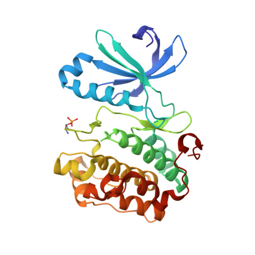
    3NUS, 3NUU, 3NUY

  • PubMed Abstract: 

    Fragment screening of phosphoinositide-dependent kinase-1 (PDK1) in a biochemical kinase assay afforded hits that were characterized and prioritized based on ligand efficiency and binding interactions with PDK1 as determined by NMR. Subsequent crystallography and follow-up screening led to the discovery of aminoindazole 19, a potent leadlike PDK1 inhibitor with high ligand efficiency. Well-defined structure-activity relationships and protein crystallography provide a basis for further elaboration and optimization of 19 as a PDK1 inhibitor.


  • Organizational Affiliation

    Oncology Research, Signal Transduction DPU Medicinal Chemistry.


Macromolecules
Find similar proteins by:  (by identity cutoff)  |  3D Structure
Entity ID: 1
MoleculeChains Sequence LengthOrganismDetailsImage
PkB-like286Homo sapiensMutation(s): 0 
Gene Names: PkB-like 1
UniProt & NIH Common Fund Data Resources
Find proteins for O15530 (Homo sapiens)
Explore O15530 
Go to UniProtKB:  O15530
PHAROS:  O15530
GTEx:  ENSG00000140992 
Entity Groups  
Sequence Clusters30% Identity50% Identity70% Identity90% Identity95% Identity100% Identity
UniProt GroupO15530
Sequence Annotations
Expand
  • Reference Sequence
Small Molecules
Ligands 3 Unique
IDChains Name / Formula / InChI Key2D Diagram3D Interactions
JOZ
Query on JOZ

Download Ideal Coordinates CCD File 
P [auth A]3,4-dihydroisoquinolin-1(2H)-one
C9 H9 N O
YWPMKTWUFVOFPL-UHFFFAOYSA-N
SO4
Query on SO4

Download Ideal Coordinates CCD File 
F [auth A]
G [auth A]
H [auth A]
I [auth A]
J [auth A]
F [auth A],
G [auth A],
H [auth A],
I [auth A],
J [auth A],
K [auth A],
L [auth A],
M [auth A],
N [auth A],
O [auth A]
SULFATE ION
O4 S
QAOWNCQODCNURD-UHFFFAOYSA-L
GOL
Query on GOL

Download Ideal Coordinates CCD File 
B [auth A],
C [auth A],
D [auth A],
E [auth A]
GLYCEROL
C3 H8 O3
PEDCQBHIVMGVHV-UHFFFAOYSA-N
Modified Residues  1 Unique
IDChains TypeFormula2D DiagramParent
SEP
Query on SEP
A
L-PEPTIDE LINKINGC3 H8 N O6 PSER
Binding Affinity Annotations 
IDSourceBinding Affinity
JOZ PDBBind:  3NUU IC50: 1.69e+5 (nM) from 1 assay(s)
BindingDB:  3NUU IC50: 1.69e+5 (nM) from 1 assay(s)
Experimental Data & Validation

Experimental Data

  • Method: X-RAY DIFFRACTION
  • Resolution: 1.98 Å
  • R-Value Free: 0.239 
  • R-Value Work: 0.189 
  • Space Group: P 32 2 1
Unit Cell:
Length ( Å )Angle ( ˚ )
a = 122.615α = 90
b = 122.615β = 90
c = 47.328γ = 120
Software Package:
Software NamePurpose
DENZOdata reduction
SCALEPACKdata scaling
PHASERphasing
PHENIXrefinement
PDB_EXTRACTdata extraction

Structure Validation

View Full Validation Report


Entry History 

Deposition Data

Revision History  (Full details and data files)

  • Version 1.0: 2011-05-25
    Type: Initial release
  • Version 1.1: 2011-07-13
    Changes: Version format compliance
  • Version 1.2: 2018-10-10
    Changes: Data collection, Database references, Structure summary