3NNW

Crystal structure of P38 alpha in complex with DP802


Experimental Data Snapshot

  • Method: X-RAY DIFFRACTION
  • Resolution: 1.89 Å
  • R-Value Free: 0.213 
  • R-Value Work: 0.167 
  • R-Value Observed: 0.169 

wwPDB Validation   3D Report Full Report


Ligand Structure Quality Assessment 


This is version 1.3 of the entry. See complete history


Literature

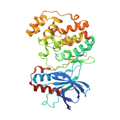
Switch control pocket inhibitors of p38-MAP kinase. Durable type II inhibitors that do not require binding into the canonical ATP hinge region

Ahn, Y.M.Clare, M.Ensinger, C.L.Hood, M.M.Lord, J.W.Lu, W.P.Miller, D.F.Patt, W.C.Smith, B.D.Vogeti, L.Kaufman, M.D.Petillo, P.A.Wise, S.C.Abendroth, J.Chun, L.Clark, R.Feese, M.Kim, H.Stewart, L.Flynn, D.L.

(2010) Bioorg Med Chem Lett 20: 5793-5798

  • DOI: https://doi.org/10.1016/j.bmcl.2010.07.134
  • Primary Citation of Related Structures:  
    3NNU, 3NNV, 3NNW, 3NNX

  • PubMed Abstract: 

    Switch control pocket inhibitors of p38-alpha kinase are described. Durable type II inhibitors were designed which bind to arginines (Arg67 or Arg70) that function as key residues for mediating phospho-threonine 180 dependant conformational fluxing of p38-alpha from an inactive type II state to an active type I state. Binding to Arg70 in particular led to potent inhibitors, exemplified by DP-802, which also exhibited high kinase selectivity. Binding to Arg70 obviated the requirement for binding into the ATP Hinge region. X-ray crystallography revealed that DP-802 and analogs induce an enhanced type II conformation upon binding to either the unphosphorylated or the doubly phosphorylated form of p38-alpha kinase.


  • Organizational Affiliation

    Deciphera Pharmaceuticals LLC, 643 Massachusetts St, Lawrence, KS 66044, USA.


Macromolecules
Find similar proteins by:  (by identity cutoff)  |  3D Structure
Entity ID: 1
MoleculeChains Sequence LengthOrganismDetailsImage
Mitogen-activated protein kinase 14354Homo sapiensMutation(s): 0 
Gene Names: MAPK14CSBPCSBP1CSBP2CSPB1MXI2
EC: 2.7.11.24
UniProt & NIH Common Fund Data Resources
Find proteins for Q16539 (Homo sapiens)
Explore Q16539 
Go to UniProtKB:  Q16539
PHAROS:  Q16539
GTEx:  ENSG00000112062 
Entity Groups  
Sequence Clusters30% Identity50% Identity70% Identity90% Identity95% Identity100% Identity
UniProt GroupQ16539
Sequence Annotations
Expand
  • Reference Sequence
Small Molecules
Ligands 1 Unique
IDChains Name / Formula / InChI Key2D Diagram3D Interactions
EDD
Query on EDD

Download Ideal Coordinates CCD File 
B [auth A]2-[3-(3-tert-butyl-5-{[(2,3-dichlorophenyl)carbamoyl]imino}-2,5-dihydro-1H-pyrazol-1-yl)phenyl]acetamide
C22 H23 Cl2 N5 O2
LLLDJNNXZHGRQO-ZXVVBBHZSA-N
Binding Affinity Annotations 
IDSourceBinding Affinity
EDD PDBBind:  3NNW IC50: 9 (nM) from 1 assay(s)
Binding MOAD:  3NNW IC50: 9 (nM) from 1 assay(s)
Experimental Data & Validation

Experimental Data

  • Method: X-RAY DIFFRACTION
  • Resolution: 1.89 Å
  • R-Value Free: 0.213 
  • R-Value Work: 0.167 
  • R-Value Observed: 0.169 
  • Space Group: P 21 21 21
Unit Cell:
Length ( Å )Angle ( ˚ )
a = 45.551α = 90
b = 85.956β = 90
c = 126.584γ = 90
Software Package:
Software NamePurpose
StructureStudiodata collection
REFMACrefinement
HKL-2000data reduction
HKL-2000data scaling

Structure Validation

View Full Validation Report


Ligand Structure Quality Assessment 


Entry History 

Deposition Data

Revision History  (Full details and data files)

  • Version 1.0: 2010-09-15
    Type: Initial release
  • Version 1.1: 2011-07-13
    Changes: Version format compliance
  • Version 1.2: 2017-11-08
    Changes: Refinement description
  • Version 1.3: 2023-12-27
    Changes: Data collection, Database references, Derived calculations