3NEV

Crystal structure of YagE, a prophage protein from E. coli K12 in complex with KDGal


Experimental Data Snapshot

  • Method: X-RAY DIFFRACTION
  • Resolution: 2.19 Å
  • R-Value Free: 0.237 
  • R-Value Work: 0.197 
  • R-Value Observed: 0.199 

wwPDB Validation   3D Report Full Report


Ligand Structure Quality Assessment 


This is version 1.2 of the entry. See complete history


Literature

Identification of biochemical and putative biological role of a xenolog from Escherichia coli using structural analysis.

Bhaskar, V.Kumar, M.Manicka, S.Tripathi, S.Venkatraman, A.Krishnaswamy, S.

(2011) Proteins 79: 1132-1142

  • DOI: https://doi.org/10.1002/prot.22949
  • Primary Citation of Related Structures:  
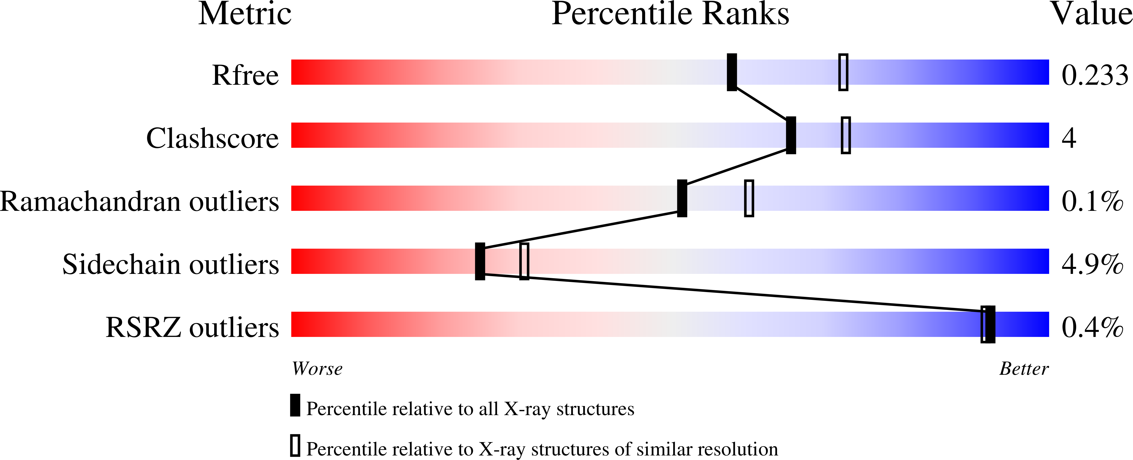
    3N2X, 3NEV

  • PubMed Abstract: 

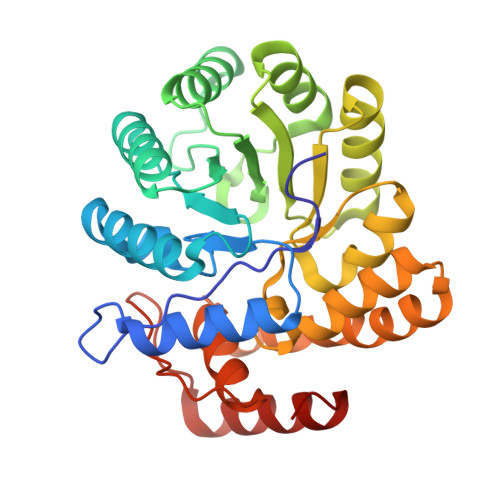
    YagE is a 33 kDa prophage protein encoded by CP4-6 prophage element in Escherichia coli K12 genome. Here, we report the structures of YagE complexes with pyruvate (PDB Id 3N2X) and KDGal (2-keto-3-deoxy galactonate) (PDB Id 3NEV) at 2.2A resolution. Pyruvate depletion assay in presence of glyceraldehyde shows that YagE catalyses the aldol condensation of pyruvate and glyceraldehyde. Our results indicate that the biochemical function of YagE is that of a 2-keto-3-deoxy gluconate (KDG) aldolase. Interestingly, E. coli K12 genome lacks an intrinsic KDG aldolase. Moreover, the over-expression of YagE increases cell viability in the presence of certain bactericidal antibiotics, indicating a putative biological role of YagE as a prophage encoded virulence factor enabling the survival of bacteria in the presence of certain antibiotics. The analysis implies a possible mechanism of antibiotic resistance conferred by the over-expression of prophage encoded YagE to E. coli.


  • Organizational Affiliation

    Department of Genetic Engineering, School of Biotechnology, Madurai Kamaraj University, Madurai 625021, India.


Macromolecules
Find similar proteins by:  (by identity cutoff)  |  3D Structure
Entity ID: 1
MoleculeChains Sequence LengthOrganismDetailsImage
Uncharacterized protein yagE
A, B, C, D
298Escherichia coli K-12Mutation(s): 0 
Gene Names: b0268JW0261yagE
UniProt
Find proteins for P75682 (Escherichia coli (strain K12))
Explore P75682 
Go to UniProtKB:  P75682
Entity Groups  
Sequence Clusters30% Identity50% Identity70% Identity90% Identity95% Identity100% Identity
UniProt GroupP75682
Sequence Annotations
Expand
  • Reference Sequence
Small Molecules
Ligands 2 Unique
IDChains Name / Formula / InChI Key2D Diagram3D Interactions
RSH
Query on RSH

Download Ideal Coordinates CCD File 
G [auth A],
J [auth B],
M [auth C],
P [auth D]
3-DEOXY-D-LYXO-HEXONIC ACID
C6 H12 O6
YGMNHEPVTNXLLS-MROZADKFSA-N
EDO
Query on EDO

Download Ideal Coordinates CCD File 
E [auth A]
F [auth A]
H [auth B]
I [auth B]
K [auth C]
E [auth A],
F [auth A],
H [auth B],
I [auth B],
K [auth C],
L [auth C],
N [auth D],
O [auth D]
1,2-ETHANEDIOL
C2 H6 O2
LYCAIKOWRPUZTN-UHFFFAOYSA-N
Experimental Data & Validation

Experimental Data

  • Method: X-RAY DIFFRACTION
  • Resolution: 2.19 Å
  • R-Value Free: 0.237 
  • R-Value Work: 0.197 
  • R-Value Observed: 0.199 
  • Space Group: P 21 21 2
Unit Cell:
Length ( Å )Angle ( ˚ )
a = 141.86α = 90
b = 155.13β = 90
c = 55.71γ = 90
Software Package:
Software NamePurpose
XSCALEdata scaling
PHASERphasing
REFMACrefinement
PDB_EXTRACTdata extraction

Structure Validation

View Full Validation Report


Ligand Structure Quality Assessment 


Entry History 

Deposition Data

Revision History  (Full details and data files)

  • Version 1.0: 2011-04-13
    Type: Initial release
  • Version 1.1: 2011-07-13
    Changes: Version format compliance
  • Version 1.2: 2023-11-01
    Changes: Data collection, Database references, Derived calculations, Refinement description