3N9V

Crystal Structure of INPP5B


Experimental Data Snapshot

  • Method: X-RAY DIFFRACTION
  • Resolution: 2.65 Å
  • R-Value Free: 0.231 
  • R-Value Work: 0.190 
  • R-Value Observed: 0.191 

wwPDB Validation   3D Report Full Report


This is version 1.3 of the entry. See complete history


Literature

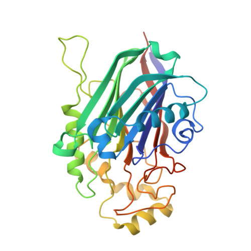
Structural basis for phosphoinositide substrate recognition, catalysis, and membrane interactions in human inositol polyphosphate 5-phosphatases

Tresaugues, L.Silvander, C.Flodin, S.Welin, M.Nyman, T.Graslund, S.Hammarstrom, M.Berglund, H.Nordlund, P.

(2014) Structure 22: 744-755

  • DOI: https://doi.org/10.1016/j.str.2014.01.013
  • Primary Citation of Related Structures:  
    3MTC, 3N9V, 3NR8, 4CML, 4CMN

  • PubMed Abstract: 

    SHIP2, OCRL, and INPP5B belong to inositol polyphosphate 5-phophatase subfamilies involved in insulin regulation and Lowes syndrome. The structural basis for membrane recognition, substrate specificity, and regulation of inositol polyphosphate 5-phophatases is still poorly understood. We determined the crystal structures of human SHIP2, OCRL, and INPP5B, the latter in complex with phosphoinositide substrate analogs, which revealed a membrane interaction patch likely to assist in sequestering substrates from the lipid bilayer. Residues recognizing the 1-phosphate of the substrates are highly conserved among human family members, suggesting similar substrate binding modes. However, 3- and 4-phosphate recognition varies and determines individual substrate specificity profiles. The high conservation of the environment of the scissile 5-phosphate suggests a common reaction geometry for all members of the human 5-phosphatase family.


  • Organizational Affiliation

    Structural Genomics Consortium, Karolinska Institutet, 17177 Stockholm, Sweden.


Macromolecules
Find similar proteins by:  (by identity cutoff)  |  3D Structure
Entity ID: 1
MoleculeChains Sequence LengthOrganismDetailsImage
Type II inositol-1,4,5-trisphosphate 5-phosphatase
A, B
313Homo sapiensMutation(s): 0 
Gene Names: INPP5B
EC: 3.1.3.36
UniProt & NIH Common Fund Data Resources
Find proteins for P32019 (Homo sapiens)
Explore P32019 
Go to UniProtKB:  P32019
PHAROS:  P32019
GTEx:  ENSG00000204084 
Entity Groups  
Sequence Clusters30% Identity50% Identity70% Identity90% Identity95% Identity100% Identity
UniProt GroupP32019
Sequence Annotations
Expand
  • Reference Sequence
Experimental Data & Validation

Experimental Data

  • Method: X-RAY DIFFRACTION
  • Resolution: 2.65 Å
  • R-Value Free: 0.231 
  • R-Value Work: 0.190 
  • R-Value Observed: 0.191 
  • Space Group: I 21 21 21
Unit Cell:
Length ( Å )Angle ( ˚ )
a = 96.16α = 90
b = 110.66β = 90
c = 159.38γ = 90
Software Package:
Software NamePurpose
ADSCdata collection
PHASERphasing
PHENIXrefinement
iMOSFLMdata reduction
SCALAdata scaling

Structure Validation

View Full Validation Report


Entry History 

Revision History  (Full details and data files)

  • Version 1.0: 2010-07-28
    Type: Initial release
  • Version 1.1: 2011-07-13
    Changes: Version format compliance
  • Version 1.2: 2014-05-28
    Changes: Database references
  • Version 1.3: 2023-09-06
    Changes: Data collection, Database references, Derived calculations, Refinement description