3N7Q

Crystal structure of human mitochondrial mTERF fragment (aa 99-399) in complex with a 12-mer DNA encompassing the tRNALeu(UUR) binding sequence


Experimental Data Snapshot

  • Method: X-RAY DIFFRACTION
  • Resolution: 2.40 Å
  • R-Value Free: 0.278 
  • R-Value Work: 0.228 
  • R-Value Observed: 0.231 

wwPDB Validation   3D Report Full Report


This is version 1.2 of the entry. See complete history


Literature

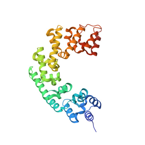
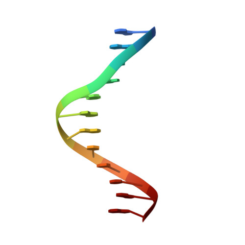
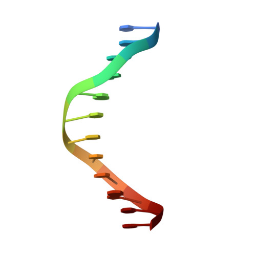
Human mitochondrial mTERF wraps around DNA through a left-handed superhelical tandem repeat.

Jimenez-Menendez, N.Fernandez-Millan, P.Rubio-Cosials, A.Arnan, C.Montoya, J.Jacobs, H.T.Bernado, P.Coll, M.Uson, I.Sola, M.

(2010) Nat Struct Mol Biol 17: 891-893

  • DOI: https://doi.org/10.1038/nsmb.1859
  • Primary Citation of Related Structures:  
    3N6S, 3N7Q

  • PubMed Abstract: 

    The regulation of mitochondrial DNA (mtDNA) processes is slowly being characterized at a structural level. We present here crystal structures of human mitochondrial regulator mTERF, a transcription termination factor also implicated in replication pausing, in complex with double-stranded DNA oligonucleotides containing the tRNA(Leu)(UUR) gene sequence. mTERF comprises nine left-handed helical tandem repeats that form a left-handed superhelix, the Zurdo domain.


  • Organizational Affiliation

    Institut de Biologia Molecular de Barcelona, Parc Cientific de Barcelona, Barcelona, Spain.


Macromolecules

Find similar proteins by:  (by identity cutoff)  |  3D Structure
Entity ID: 1
MoleculeChains Sequence LengthOrganismDetailsImage
Transcription termination factor, mitochondrial310Homo sapiensMutation(s): 0 
Gene Names: MTERF
UniProt & NIH Common Fund Data Resources
Find proteins for Q99551 (Homo sapiens)
Explore Q99551 
Go to UniProtKB:  Q99551
GTEx:  ENSG00000127989 
Entity Groups  
Sequence Clusters30% Identity50% Identity70% Identity90% Identity95% Identity100% Identity
UniProt GroupQ99551
Sequence Annotations
Expand
  • Reference Sequence

Find similar nucleic acids by:  Sequence   |   3D Structure  

Entity ID: 2
MoleculeChains LengthOrganismImage
DNA (5'-D(*GP*GP*CP*AP*GP*AP*GP*CP*CP*CP*GP*G)-3')12N/A
Sequence Annotations
Expand
  • Reference Sequence

Find similar nucleic acids by:  Sequence   |   3D Structure  

Entity ID: 3
MoleculeChains LengthOrganismImage
DNA (5'-D(*CP*CP*GP*GP*GP*CP*TP*CP*TP*GP*CP*C)-3')12N/A
Sequence Annotations
Expand
  • Reference Sequence
Small Molecules
Ligands 1 Unique
IDChains Name / Formula / InChI Key2D Diagram3D Interactions
CIT
Query on CIT

Download Ideal Coordinates CCD File 
D [auth A]CITRIC ACID
C6 H8 O7
KRKNYBCHXYNGOX-UHFFFAOYSA-N
Experimental Data & Validation

Experimental Data

  • Method: X-RAY DIFFRACTION
  • Resolution: 2.40 Å
  • R-Value Free: 0.278 
  • R-Value Work: 0.228 
  • R-Value Observed: 0.231 
  • Space Group: P 32
Unit Cell:
Length ( Å )Angle ( ˚ )
a = 99.851α = 90
b = 99.851β = 90
c = 39.6γ = 120
Software Package:
Software NamePurpose
EPMRphasing
PHENIXrefinement
XDSdata reduction
SHELXdata scaling

Structure Validation

View Full Validation Report


Entry History 

Deposition Data

Revision History  (Full details and data files)

  • Version 1.0: 2010-06-16
    Type: Initial release
  • Version 1.1: 2011-07-13
    Changes: Version format compliance
  • Version 1.2: 2024-02-21
    Changes: Data collection, Database references, Derived calculations