Structure of the novel 14kDa fragment of alpha-subunit of phycoerythrin from the starving cyanobacterium Phormidium tenue.
Soni, B.R., Hasan, M.I., Parmar, A., Ethayathulla, A.S., Kumar, R.P., Singh, N.K., Sinha, M., Kaur, P., Yadav, S., Sharma, S., Madamwar, D., Singh, T.P.(2010) J Struct Biol 171: 247-255
- PubMed: 20546902
- DOI: https://doi.org/10.1016/j.jsb.2010.05.008
- Primary Citation of Related Structures:
3MWN - PubMed Abstract:
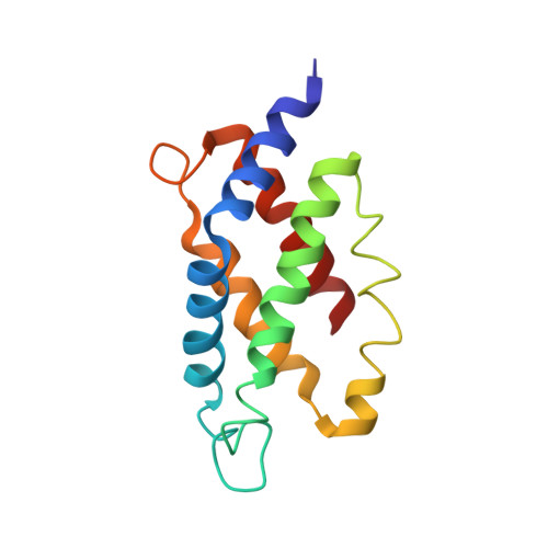
The rod-like phycobilisome (PBS) in cyanobacterium is the light-harvesting complex of phycoerythrin (PE), phycocyanin (PC) and allophycocyanin (APC). The orderly degradation of PBS was observed under starvation conditions. A 14 kDa truncated fragment of alpha-subunit of PE (F-alphaPE) was identified from the degraded product. F-alphaPE was purified to homogeneity, sequenced and crystallized. The merohedrally twinned crystals with a twinning factor of approximately 0.5 were obtained. The crystal structure of F-alphaPE was determined with molecular replacement method using detwinned data and refined to an R(cryst) factor of 23.2% (R(free)=27.6%). The structure consisted of two crystallographically independent molecules in the asymmetric unit. The two molecules were designated as molecules A and B with a buried area of 200 A(2) at the interface. The structure of F-alphaPE consists of seven alpha-helices A, B, E, F, F', G and H. The first 31N-terminal residues that fold into parallel alpha-helices X and Y in other PEs are not present in the amino acid sequence of F-alphaPE. Both molecules, A and B contain two chromophore ligands, PEB1 and PEB2 in each. These are covalently linked to the polypeptide chain through Cys82 and Cys139, respectively. The superimposition of C(alpha) tracings of molecules A and B shows an r.m.s. shift of 1.0 A indicating that the structures of two independent molecules are very similar. The degradation of phycobilisome proteins under starvation stress seems to occur to supplement the requirement of amino acids for protein synthesis and to reduce the absorption of light energy.
Organizational Affiliation:
BRD School of Biosciences, Sardar Patel University, Vallabh Vidyanagar 388120, India.