3MW2

Crystal structure of beta-neurexin 1 with the splice insert 4


Experimental Data Snapshot

  • Method: X-RAY DIFFRACTION
  • Resolution: 2.69 Å
  • R-Value Free: 0.287 
  • R-Value Work: 0.206 
  • R-Value Observed: 0.210 

wwPDB Validation   3D Report Full Report


This is version 2.1 of the entry. See complete history


Literature

Splice Form Dependence of beta-Neurexin/Neuroligin Binding Interactions.

Koehnke, J.Katsamba, P.S.Ahlsen, G.Bahna, F.Vendome, J.Honig, B.Shapiro, L.Jin, X.

(2010) Neuron 67: 61-74

  • DOI: https://doi.org/10.1016/j.neuron.2010.06.001
  • Primary Citation of Related Structures:  
    3MW2, 3MW3, 3MW4

  • PubMed Abstract: 

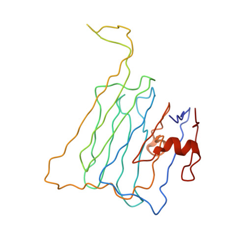
    Alternatively spliced beta-neurexins (beta-NRXs) and neuroligins (NLs) are thought to have distinct extracellular binding affinities, potentially providing a beta-NRX/NL synaptic recognition code. We utilized surface plasmon resonance to measure binding affinities between all combinations of alternatively spliced beta-NRX 1-3 and NL 1-3 ectodomains. Binding was observed for all beta-NRX/NL pairs. The presence of the NL1 B splice insertion lowers beta-NRX binding affinity by approximately 2-fold, while beta-NRX splice insertion 4 has small effects that do not synergize with NL splicing. New structures of glycosylated beta-NRXs 1 and 2 containing splice insertion 4 reveal that the insertion forms a new beta strand that replaces the beta10 strand, leaving the NL binding site intact. This helps to explain the limited effect of splice insert 4 on NRX/NL binding affinities. These results provide new structural insights and quantitative binding information to help determine whether and how splice isoform choice plays a role in beta-NRX/NL-mediated synaptic recognition.


  • Organizational Affiliation

    Department of Biochemistry and Molecular Biophysics, Columbia University, New York, NY 10032, USA.


Macromolecules
Find similar proteins by:  (by identity cutoff)  |  3D Structure
Entity ID: 1
MoleculeChains Sequence LengthOrganismDetailsImage
Neurexin-1-alpha
A, B
207Mus musculusMutation(s): 0 
Gene Names: Nrxn1Kiaa0578
UniProt & NIH Common Fund Data Resources
Find proteins for Q9CS84 (Mus musculus)
Explore Q9CS84 
Go to UniProtKB:  Q9CS84
IMPC:  MGI:1096391
Entity Groups  
Sequence Clusters30% Identity50% Identity70% Identity90% Identity95% Identity100% Identity
UniProt GroupQ9CS84
Sequence Annotations
Expand
  • Reference Sequence
Oligosaccharides

Help

Entity ID: 2
MoleculeChains Length2D Diagram Glycosylation3D Interactions
2-acetamido-2-deoxy-beta-D-glucopyranose-(1-4)-[beta-D-mannopyranose-(1-6)]2-acetamido-2-deoxy-beta-D-glucopyranose
C, D
3N-Glycosylation
Glycosylation Resources
GlyTouCan:  G16421CZ
GlyCosmos:  G16421CZ
GlyGen:  G16421CZ
Small Molecules
Ligands 1 Unique
IDChains Name / Formula / InChI Key2D Diagram3D Interactions
PO4
Query on PO4

Download Ideal Coordinates CCD File 
E [auth A]PHOSPHATE ION
O4 P
NBIIXXVUZAFLBC-UHFFFAOYSA-K
Experimental Data & Validation

Experimental Data

  • Method: X-RAY DIFFRACTION
  • Resolution: 2.69 Å
  • R-Value Free: 0.287 
  • R-Value Work: 0.206 
  • R-Value Observed: 0.210 
  • Space Group: C 1 2 1
Unit Cell:
Length ( Å )Angle ( ˚ )
a = 85.89α = 90
b = 59.819β = 103.86
c = 105.697γ = 90
Software Package:
Software NamePurpose
MAR345data collection
PHASERphasing
REFMACrefinement
HKL-2000data reduction
SCALEPACKdata scaling

Structure Validation

View Full Validation Report


Entry History 

Deposition Data

Revision History  (Full details and data files)

  • Version 1.0: 2010-07-28
    Type: Initial release
  • Version 1.1: 2011-07-13
    Changes: Advisory, Version format compliance
  • Version 1.2: 2017-11-08
    Changes: Refinement description
  • Version 2.0: 2020-07-29
    Type: Remediation
    Reason: Carbohydrate remediation
    Changes: Atomic model, Data collection, Database references, Derived calculations, Structure summary
  • Version 2.1: 2023-09-06
    Changes: Data collection, Database references, Refinement description, Structure summary