3MVS

Structure of the N-terminus of Cadherin 23


Experimental Data Snapshot

  • Method: X-RAY DIFFRACTION
  • Resolution: 1.10 Å
  • R-Value Free: 0.188 
  • R-Value Work: 0.163 
  • R-Value Observed: 0.164 

wwPDB Validation   3D Report Full Report


This is version 1.2 of the entry. See complete history


Literature

Structure of the N terminus of cadherin 23 reveals a new adhesion mechanism for a subset of cadherin superfamily members.

Elledge, H.M.Kazmierczak, P.Clark, P.Joseph, J.S.Kolatkar, A.Kuhn, P.Muller, U.

(2010) Proc Natl Acad Sci U S A 107: 10708-10712

  • DOI: https://doi.org/10.1073/pnas.1006284107
  • Primary Citation of Related Structures:  
    3MVS

  • PubMed Abstract: 

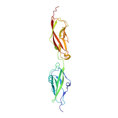
    The cadherin superfamily encodes more than 100 receptors with diverse functions in tissue development and homeostasis. Classical cadherins mediate adhesion by binding interactions that depend on their N-terminal extracellular cadherin (EC) domains, which swap N-terminal beta-strands. Sequence alignments suggest that the strand-swap binding mode is not commonly used by functionally divergent cadherins. Here, we have determined the structure of the EC1-EC2 domains of cadherin 23 (CDH23), which binds to protocadherin 15 (PCDH15) to form tip links of mechanosensory hair cells. Unlike classical cadherins, the CDH23 N terminus contains polar amino acids that bind Ca(2+). The N terminus of PCDH15 also contains polar amino acids. Mutations in polar amino acids within EC1 of CDH23 and PCDH15 abolish interaction between the two cadherins. PCDH21 and PCDH24 contain similarly charged N termini, suggesting that a subset of cadherins share a common interaction mechanism that differs from the strand-swap binding mode of classical cadherins.


  • Organizational Affiliation

    Department of Cell Biology and Dorris Neuroscience Center, The Scripps Research Institute, La Jolla, CA 92037, USA.


Macromolecules
Find similar proteins by:  (by identity cutoff)  |  3D Structure
Entity ID: 1
MoleculeChains Sequence LengthOrganismDetailsImage
Cadherin-23210Mus musculusMutation(s): 0 
Gene Names: Cdh23
UniProt & NIH Common Fund Data Resources
Find proteins for Q99PF4 (Mus musculus)
Explore Q99PF4 
Go to UniProtKB:  Q99PF4
IMPC:  MGI:1890219
Entity Groups  
Sequence Clusters30% Identity50% Identity70% Identity90% Identity95% Identity100% Identity
UniProt GroupQ99PF4
Sequence Annotations
Expand
  • Reference Sequence
Small Molecules
Ligands 2 Unique
IDChains Name / Formula / InChI Key2D Diagram3D Interactions
EDO
Query on EDO

Download Ideal Coordinates CCD File 
AA [auth A]
BA [auth A]
CA [auth A]
H [auth A]
I [auth A]
AA [auth A],
BA [auth A],
CA [auth A],
H [auth A],
I [auth A],
J [auth A],
K [auth A],
L [auth A],
M [auth A],
N [auth A],
O [auth A],
P [auth A],
Q [auth A],
R [auth A],
S [auth A],
T [auth A],
U [auth A],
V [auth A],
W [auth A],
X [auth A],
Y [auth A],
Z [auth A]
1,2-ETHANEDIOL
C2 H6 O2
LYCAIKOWRPUZTN-UHFFFAOYSA-N
CA
Query on CA

Download Ideal Coordinates CCD File 
B [auth A]
C [auth A]
D [auth A]
E [auth A]
F [auth A]
B [auth A],
C [auth A],
D [auth A],
E [auth A],
F [auth A],
G [auth A]
CALCIUM ION
Ca
BHPQYMZQTOCNFJ-UHFFFAOYSA-N
Experimental Data & Validation

Experimental Data

  • Method: X-RAY DIFFRACTION
  • Resolution: 1.10 Å
  • R-Value Free: 0.188 
  • R-Value Work: 0.163 
  • R-Value Observed: 0.164 
  • Space Group: P 1 21 1
Unit Cell:
Length ( Å )Angle ( ˚ )
a = 38.375α = 90
b = 64.208β = 110.89
c = 47.859γ = 90
Software Package:
Software NamePurpose
HKL-2000data collection
PHASERphasing
REFMACrefinement
HKL-2000data reduction
HKL-2000data scaling

Structure Validation

View Full Validation Report


Entry History 

Deposition Data

Revision History  (Full details and data files)

  • Version 1.0: 2010-06-09
    Type: Initial release
  • Version 1.1: 2011-07-13
    Changes: Version format compliance
  • Version 1.2: 2024-02-21
    Changes: Data collection, Database references, Derived calculations