


The Crystal Structure of a putative 4-hydroxy-2-oxoglutarate aldolase from Bacillus anthracis to 1.45A
Stein, A.J., Hatzos-Skintges, C., Clancy, S., Joachimiak, A.To be published.
Experimental Data Snapshot
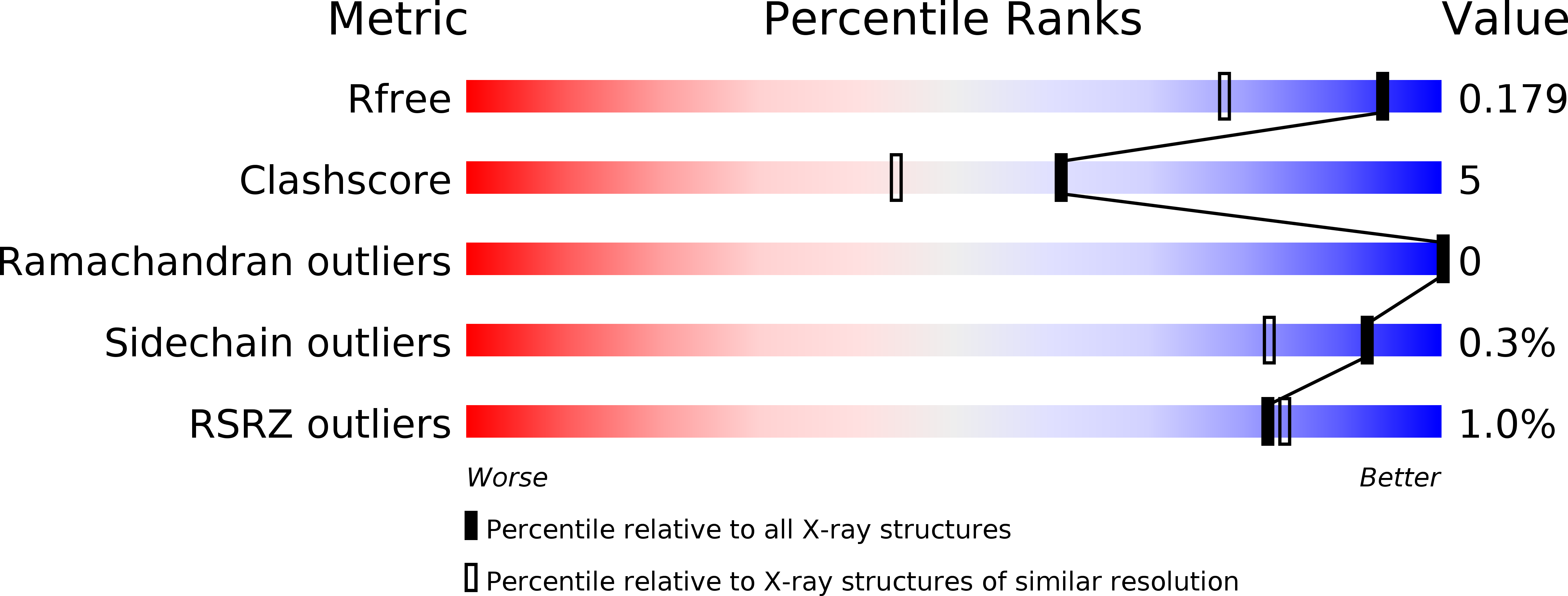
wwPDB Validation 3D Report Full Report
Entity ID: 1 | |||||
|---|---|---|---|---|---|
| Molecule | Chains | Sequence Length | Organism | Details | Image |
| putative 4-hydroxy-2-oxoglutarate aldolase | 251 | Bacillus anthracis str. Sterne | Mutation(s): 0 Gene Names: BAS4738, BA_5098, GBAA5098, GBAA_5098 | 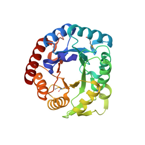 | |
UniProt | |||||
Find proteins for A0A6L7GXV7 (Bacillus anthracis) Explore A0A6L7GXV7 Go to UniProtKB: A0A6L7GXV7 | |||||
Entity Groups | |||||
| Sequence Clusters | 30% Identity50% Identity70% Identity90% Identity95% Identity100% Identity | ||||
| UniProt Group | A0A6L7GXV7 | ||||
Sequence AnnotationsExpand | |||||
| |||||
| Ligands 2 Unique | |||||
|---|---|---|---|---|---|
| ID | Chains | Name / Formula / InChI Key | 2D Diagram | 3D Interactions | |
| CL Query on CL | C [auth A], E [auth B], F [auth B] | CHLORIDE ION Cl VEXZGXHMUGYJMC-UHFFFAOYSA-M |  | ||
| NA Query on NA | D [auth A], G [auth B] | SODIUM ION Na FKNQFGJONOIPTF-UHFFFAOYSA-N |  | ||
| Modified Residues 1 Unique | |||||
|---|---|---|---|---|---|
| ID | Chains | Type | Formula | 2D Diagram | Parent |
| MSE Query on MSE | A, B | L-PEPTIDE LINKING | C5 H11 N O2 Se |  | MET |
| Length ( Å ) | Angle ( ˚ ) |
|---|---|
| a = 55.669 | α = 90 |
| b = 68.431 | β = 90 |
| c = 131.081 | γ = 90 |
| Software Name | Purpose |
|---|---|
| DENZO | data reduction |
| SCALEPACK | data scaling |
| REFMAC | refinement |
| PDB_EXTRACT | data extraction |
| SBC-Collect | data collection |
| HKL-3000 | data reduction |
| HKL-3000 | data scaling |
| SHELX | phasing |
| MLPHARE | phasing |
| DM | phasing |
| ARP/wARP | model building |
| Coot | model building |
RCSB PDB (citation) is hosted by
RCSB PDB is a member of the

