Modulation of inhibitory activity of xylanase-alpha-amylase inhibitor protein (XAIP): binding studies and crystal structure determination of XAIP-II from Scadoxus multiflorus at 1.2 A resolution.
Kumar, S., Singh, N., Mishra, B., Dube, D., Sinha, M., Singh, S.B., Dey, S., Kaur, P., Sharma, S., Singh, T.P.(2010) BMC Struct Biol 10: 41-41
- PubMed: 21092126
- DOI: https://doi.org/10.1186/1472-6807-10-41
- Primary Citation of Related Structures:
3MU7 - PubMed Abstract:
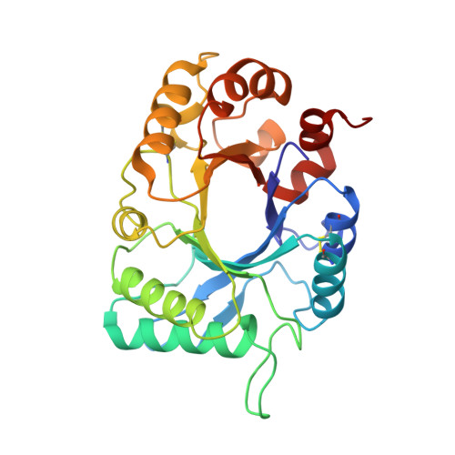
Plants produce a wide range of proteinaceous inhibitors to protect themselves against hydrolytic enzymes. Recently a novel protein XAIP belonging to a new sub-family (GH18C) was reported to inhibit two structurally unrelated enzymes xylanase GH11 and α-amylase GH13. It was shown to inhibit xylanase GH11 with greater potency than that of α-amylase GH13. A new form of XAIP (XAIP-II) that inhibits α-amylase GH13 with a greater potency than that of XAIP and xylanase GH11 with a lower potency than that of XAIP, has been identified in the extracts of underground bulbs of Scadoxus multiflorus. This kind of occurrence of isoforms of inhibitor proteins is a rare observation and offers new opportunities for understanding the principles of protein engineering by nature. In order to determine the structural basis of the enhanced potency of XAIP-II against α-amylase GH13 and its reduced potency against xylanase GH11 as compared to that of XAIP, we have purified XAIP-II to homogeneity and obtained its complete amino acid sequence using cloning procedure. It has been crystallized with 0.1 M ammonium sulphate as the precipitating agent and the three-dimensional structure has been determined at 1.2 Å resolution. The binding studies of XAIP-II with xylanase GH11 and α-amylase GH13 have been carried out with surface plasmon resonance (SPR). The structure determination revealed that XAIP-II adopts the well known TIM barrel fold. The xylanase GH11 binding site in XAIP-II is formed mainly with loop α3-β3 (residues, 102 - 118) which has acquired a stereochemically less favorable conformation for binding to xylanase GH11 because of the addition of an extra residue, Ala105 and due to replacements of two important residues, His106 and Asn109 by Thr107 and Ser110. On the other hand, the α-amylase binding site, which consists of α-helices α6 (residues, 193 - 206), α7 (residues, 230 - 243) and loop β6-α6 (residues, 180 - 192) adopts a stereochemically more favorable conformation due to replacements of residues, Ser190, Gly191 and Glu194 by Ala191, Ser192 and Ser195 respectively in α-helix α6, Glu231 and His236 by Thr232 and Ser237 respectively in α-helix α7. As a result, XAIP-II binds to xylanase GH11 less favorably while it interacts more strongly with α-amylase GH13 as compared to XAIP. These observations correlate well with the values of 4.2 × 10(-6) M and 3.4 × 10(-8) M for the dissociation constants of XAIP-II with xylanase GH11 and α-amylase GH13 respectively and those of 4.5 × 10(-7) M and 3.6 × 10(-6) M of XAIP with xylanase GH11 and α-amylase GH13 respectively.
Organizational Affiliation:
Department of Biophysics, All India Institute of Medical Sciences, New Delhi, India.