3MNV

Crystal structure of the non-neutralizing HIV antibody 13H11 Fab fragment


Experimental Data Snapshot

  • Method: X-RAY DIFFRACTION
  • Resolution: 2.40 Å
  • R-Value Free: 0.228 
  • R-Value Work: 0.181 
  • R-Value Observed: 0.183 

wwPDB Validation   3D Report Full Report


This is version 1.3 of the entry. See complete history


Literature

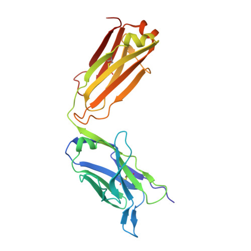
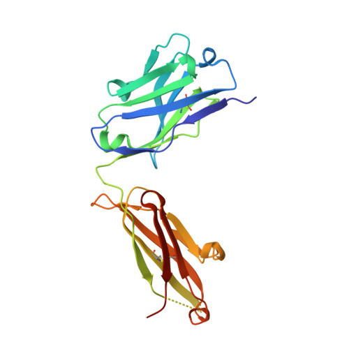
Crystal structure of a non-neutralizing antibody to the HIV-1 gp41 membrane-proximal external region.

Nicely, N.I.Dennison, S.M.Spicer, L.Scearce, R.M.Kelsoe, G.Ueda, Y.Chen, H.Liao, H.X.Alam, S.M.Haynes, B.F.

(2010) Nat Struct Mol Biol 17: 1492-1494

  • DOI: https://doi.org/10.1038/nsmb.1944
  • Primary Citation of Related Structures:  
    3MNV

  • PubMed Abstract: 

    The monoclonal antibody 13H11 shares part of its epitope in the HIV-1 gp41 membrane-proximal external region (MPER) with the rare, broadly neutralizing human antibody 2F5. Although 13H11 partially cross-blocked 2F5 binding, 13H11 is non-neutralizing and does not block 2F5 neutralization. We show that unlike 2F5, 13H11 binds to a well-defined helical MPER structure that is consistent with the structure of gp41 in a post-fusion six-helix bundle conformation.


  • Organizational Affiliation

    Duke Human Vaccine Institute, Duke University School of Medicine, Durham, North Carolina, USA. nathan.nicely@duke.edu


Macromolecules
Find similar proteins by:  (by identity cutoff)  |  3D Structure
Entity ID: 1
MoleculeChains Sequence LengthOrganismDetailsImage
ANTI-HIV-1 ANTIBODY 13H11 LIGHT CHAIN
A, C
224Mus musculusHomo sapiens
This entity is chimeric
Mutation(s): 0 
UniProt
Find proteins for Q8TCD0 (Homo sapiens)
Explore Q8TCD0 
Go to UniProtKB:  Q8TCD0
Entity Groups  
Sequence Clusters30% Identity50% Identity70% Identity90% Identity95% Identity100% Identity
UniProt GroupQ8TCD0
Sequence Annotations
Expand
  • Reference Sequence
Find similar proteins by:  (by identity cutoff)  |  3D Structure
Entity ID: 2
MoleculeChains Sequence LengthOrganismDetailsImage
ANTI-HIV-1 ANTIBODY 13H11 HEAVY CHAIN
B, D
219Mus musculusHomo sapiens
This entity is chimeric
Mutation(s): 0 
UniProt
Find proteins for Q6N089 (Homo sapiens)
Explore Q6N089 
Go to UniProtKB:  Q6N089
Entity Groups  
Sequence Clusters30% Identity50% Identity70% Identity90% Identity95% Identity100% Identity
UniProt GroupQ6N089
Sequence Annotations
Expand
  • Reference Sequence
Small Molecules
Ligands 4 Unique
IDChains Name / Formula / InChI Key2D Diagram3D Interactions
SO4
Query on SO4

Download Ideal Coordinates CCD File 
E [auth A]
F [auth B]
G [auth B]
H [auth B]
M [auth C]
E [auth A],
F [auth B],
G [auth B],
H [auth B],
M [auth C],
N [auth C]
SULFATE ION
O4 S
QAOWNCQODCNURD-UHFFFAOYSA-L
EDO
Query on EDO

Download Ideal Coordinates CCD File 
I [auth B],
J [auth B],
K [auth B],
O [auth C]
1,2-ETHANEDIOL
C2 H6 O2
LYCAIKOWRPUZTN-UHFFFAOYSA-N
NO3
Query on NO3

Download Ideal Coordinates CCD File 
P [auth C]NITRATE ION
N O3
NHNBFGGVMKEFGY-UHFFFAOYSA-N
CL
Query on CL

Download Ideal Coordinates CCD File 
L [auth B]CHLORIDE ION
Cl
VEXZGXHMUGYJMC-UHFFFAOYSA-M
Experimental Data & Validation

Experimental Data

  • Method: X-RAY DIFFRACTION
  • Resolution: 2.40 Å
  • R-Value Free: 0.228 
  • R-Value Work: 0.181 
  • R-Value Observed: 0.183 
  • Space Group: P 61
Unit Cell:
Length ( Å )Angle ( ˚ )
a = 158.378α = 90
b = 158.378β = 90
c = 92.975γ = 120
Software Package:
Software NamePurpose
PHENIXmodel building
PHENIXrefinement
d*TREKdata reduction
d*TREKdata scaling
PHENIXphasing

Structure Validation

View Full Validation Report


Entry History 

Deposition Data

Revision History  (Full details and data files)

  • Version 1.0: 2010-11-17
    Type: Initial release
  • Version 1.1: 2011-07-13
    Changes: Version format compliance
  • Version 1.2: 2017-08-16
    Changes: Source and taxonomy
  • Version 1.3: 2023-09-06
    Changes: Data collection, Database references, Derived calculations, Refinement description