3MJH

Crystal Structure of Human Rab5A in complex with the C2H2 Zinc Finger of EEA1


Experimental Data Snapshot

  • Method: X-RAY DIFFRACTION
  • Resolution: 2.03 Å
  • R-Value Free: 0.260 
  • R-Value Work: 0.192 
  • R-Value Observed: 0.196 

wwPDB Validation   3D Report Full Report


This is version 1.3 of the entry. See complete history


Literature

Structural basis for Rab GTPase recognition and endosome tethering by the C2H2 zinc finger of Early Endosomal Autoantigen 1 (EEA1).

Mishra, A.Eathiraj, S.Corvera, S.Lambright, D.G.

(2010) Proc Natl Acad Sci U S A 107: 10866-10871

  • DOI: https://doi.org/10.1073/pnas.1000843107
  • Primary Citation of Related Structures:  
    3MJH

  • PubMed Abstract: 

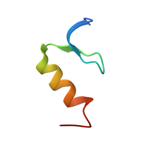
    Regulation of endosomal trafficking by Rab GTPases depends on selective interactions with multivalent effectors, including EEA1 and Rabenosyn-5, which facilitate endosome tethering, sorting, and fusion. Both EEA1 and Rabenosyn-5 contain a distinctive N-terminal C(2)H(2) zinc finger that binds Rab5. How these C(2)H(2) zinc fingers recognize Rab GTPases remains unknown. Here, we report the crystal structure of Rab5A in complex with the EEA1 C(2)H(2) zinc finger. The binding interface involves all elements of the zinc finger as well as a short N-terminal extension but is restricted to the switch and interswitch regions of Rab5. High selectivity for Rab5 and, to a lesser extent Rab22, is observed in quantitative profiles of binding to Rab family GTPases. Although critical determinants are identified in both switch regions, Rab4-to-Rab5 conversion-of-specificity mutants reveal an essential requirement for additional substitutions in the proximal protein core that are predicted to indirectly influence recognition through affects on the structure and conformational stability of the switch regions.


  • Organizational Affiliation

    Department of Biochemistry and Molecular Pharmacology, University of Massachusetts Medical School, Worcester, MA 01605, USA.


Macromolecules
Find similar proteins by:  (by identity cutoff)  |  3D Structure
Entity ID: 1
MoleculeChains Sequence LengthOrganismDetailsImage
Ras-related protein Rab-5A
A, C
168Homo sapiensMutation(s): 1 
Gene Names: RAB5RAB5A
EC: 3.6.5.2
UniProt & NIH Common Fund Data Resources
Find proteins for P20339 (Homo sapiens)
Explore P20339 
Go to UniProtKB:  P20339
PHAROS:  P20339
GTEx:  ENSG00000144566 
Entity Groups  
Sequence Clusters30% Identity50% Identity70% Identity90% Identity95% Identity100% Identity
UniProt GroupP20339
Sequence Annotations
Expand
  • Reference Sequence
Find similar proteins by:  (by identity cutoff)  |  3D Structure
Entity ID: 2
MoleculeChains Sequence LengthOrganismDetailsImage
Early endosome antigen 1
B, D
34Homo sapiensMutation(s): 0 
Gene Names: Early Endosomal Antigen1(EEA1)EEA1ZFYVE2
UniProt & NIH Common Fund Data Resources
Find proteins for Q15075 (Homo sapiens)
Explore Q15075 
Go to UniProtKB:  Q15075
PHAROS:  Q15075
GTEx:  ENSG00000102189 
Entity Groups  
Sequence Clusters30% Identity50% Identity70% Identity90% Identity95% Identity100% Identity
UniProt GroupQ15075
Sequence Annotations
Expand
  • Reference Sequence
Experimental Data & Validation

Experimental Data

  • Method: X-RAY DIFFRACTION
  • Resolution: 2.03 Å
  • R-Value Free: 0.260 
  • R-Value Work: 0.192 
  • R-Value Observed: 0.196 
  • Space Group: P 21 21 21
Unit Cell:
Length ( Å )Angle ( ˚ )
a = 46.412α = 90
b = 80.398β = 90
c = 103.495γ = 90
Software Package:
Software NamePurpose
HKL-2000data collection
AMoREphasing
REFMACrefinement
DENZOdata reduction
SCALEPACKdata scaling

Structure Validation

View Full Validation Report


Entry History 

Deposition Data

Revision History  (Full details and data files)

  • Version 1.0: 2010-05-05
    Type: Initial release
  • Version 1.1: 2011-07-13
    Changes: Version format compliance
  • Version 1.2: 2021-10-06
    Changes: Database references, Derived calculations
  • Version 1.3: 2024-02-21
    Changes: Data collection