3MJ9

Crystal structure of JAML in complex with the stimulatory antibody HL4E10


Experimental Data Snapshot

  • Method: X-RAY DIFFRACTION
  • Resolution: 2.95 Å
  • R-Value Free: 0.287 
  • R-Value Work: 0.222 
  • R-Value Observed: 0.225 

wwPDB Validation   3D Report Full Report


Ligand Structure Quality Assessment 


This is version 2.0 of the entry. See complete history


Literature

Molecular insights into gamma delta T cell costimulation by an anti-JAML antibody.

Verdino, P.Witherden, D.A.Ferguson, M.S.Corper, A.L.Schiefner, A.Havran, W.L.Wilson, I.A.

(2011) Structure 19: 80-89

  • DOI: https://doi.org/10.1016/j.str.2010.10.007
  • Primary Citation of Related Structures:  
    3MJ9

  • PubMed Abstract: 

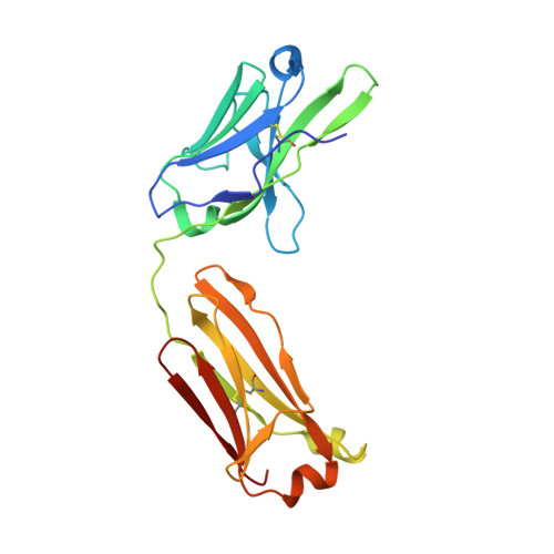
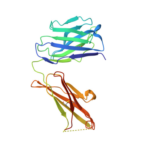
    γδ T cells bridge innate and adaptive immunity and function in immunosurveillance, immunoregulation, tumor cell recognition, and as first line of defense against microbial infection. Costimulation of epithelial γδ T cell activation by the JAML receptor can be induced by interaction with its endogenous ligand CAR or by binding of the stimulatory antibody HL4E10. We, therefore, determined the crystal structure of the JAML-HL4E10 Fab complex at 2.95 Å resolution. HL4E10 binds the membrane-proximal domain of JAML through hydrophobic interactions that account for nanomolar affinity and long half-life, contrasting with the fast kinetics and micromolar affinity of the hydrophilic CAR interaction with the membrane-distal JAML domain. Thus, despite different binding sites and mechanisms, JAML interaction with these two disparate ligands leads to the same functional outcome, namely JAML triggering and induction of cell signaling. Several characteristics of the HL4E10 antibody might then be harnessed in therapeutic applications, such as promoting healing of acute or chronic wounds.


  • Organizational Affiliation

    Department of Molecular Biology, The Scripps Research Institute, 10550 North Torrey Pines Road, La Jolla, CA 92037, USA.


Macromolecules
Find similar proteins by:  (by identity cutoff)  |  3D Structure
Entity ID: 1
MoleculeChains Sequence LengthOrganismDetailsImage
Junctional adhesion molecule-like268Mus musculusMutation(s): 2 
Gene Names: Amica1Gm638Jaml
UniProt
Find proteins for Q80UL9 (Mus musculus)
Explore Q80UL9 
Go to UniProtKB:  Q80UL9
Entity Groups  
Sequence Clusters30% Identity50% Identity70% Identity90% Identity95% Identity100% Identity
UniProt GroupQ80UL9
Sequence Annotations
Expand
  • Reference Sequence
Find similar proteins by:  (by identity cutoff)  |  3D Structure
Entity ID: 2
MoleculeChains Sequence LengthOrganismDetailsImage
STIMULATORY HAMSTER ANTIBODY HL4E10 FAB LIGHT CHAINB [auth L]213Cricetulus migratoriusMutation(s): 0 
Entity Groups  
Sequence Clusters30% Identity50% Identity70% Identity90% Identity95% Identity100% Identity
Sequence Annotations
Expand
  • Reference Sequence
Find similar proteins by:  (by identity cutoff)  |  3D Structure
Entity ID: 3
MoleculeChains Sequence LengthOrganismDetailsImage
STIMULATORY HAMSTER ANTIBODY HL4E10 FAB HEAVY CHAINC [auth H]223Cricetulus migratoriusMutation(s): 0 
Entity Groups  
Sequence Clusters30% Identity50% Identity70% Identity90% Identity95% Identity100% Identity
Sequence Annotations
Expand
  • Reference Sequence
Oligosaccharides

Help

Entity ID: 4
MoleculeChains Length2D Diagram Glycosylation3D Interactions
alpha-D-mannopyranose-(1-3)-[alpha-D-mannopyranose-(1-6)]beta-D-mannopyranose-(1-4)-2-acetamido-2-deoxy-beta-D-glucopyranose-(1-4)-[alpha-L-fucopyranose-(1-6)]2-acetamido-2-deoxy-beta-D-glucopyranoseD [auth B]6N-Glycosylation
Glycosylation Resources
GlyTouCan:  G82348BZ
GlyCosmos:  G82348BZ
GlyGen:  G82348BZ
Small Molecules
Ligands 1 Unique
IDChains Name / Formula / InChI Key2D Diagram3D Interactions
NAG
Query on NAG

Download Ideal Coordinates CCD File 
E [auth A],
F [auth A]
2-acetamido-2-deoxy-beta-D-glucopyranose
C8 H15 N O6
OVRNDRQMDRJTHS-FMDGEEDCSA-N
Experimental Data & Validation

Experimental Data

  • Method: X-RAY DIFFRACTION
  • Resolution: 2.95 Å
  • R-Value Free: 0.287 
  • R-Value Work: 0.222 
  • R-Value Observed: 0.225 
  • Space Group: P 43 21 2
Unit Cell:
Length ( Å )Angle ( ˚ )
a = 125.02α = 90
b = 125.02β = 90
c = 107.839γ = 90
Software Package:
Software NamePurpose
PHASERphasing
REFMACrefinement
HKL-2000data reduction
HKL-2000data scaling

Structure Validation

View Full Validation Report


Ligand Structure Quality Assessment 


Entry History 

Deposition Data

Revision History  (Full details and data files)

  • Version 1.0: 2011-02-23
    Type: Initial release
  • Version 1.1: 2011-07-13
    Changes: Version format compliance
  • Version 2.0: 2020-07-29
    Type: Remediation
    Reason: Carbohydrate remediation
    Changes: Atomic model, Data collection, Database references, Derived calculations, Structure summary