3MIU

Structure of Banana Lectin-pentamannose complex


Experimental Data Snapshot

  • Method: X-RAY DIFFRACTION
  • Resolution: 2.63 Å
  • R-Value Free: 0.243 
  • R-Value Work: 0.217 
  • R-Value Observed: 0.219 

wwPDB Validation   3D Report Full Report


Ligand Structure Quality Assessment 


This is version 2.1 of the entry. See complete history


Literature

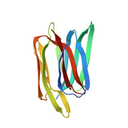
Influence of glycosidic linkage on the nature of carbohydrate binding in beta-prism I fold lectins: an X-ray and molecular dynamics investigation on banana lectin-carbohydrate complexes

Sharma, A.Vijayan, M.

(2011) Glycobiology 21: 23-33

  • DOI: https://doi.org/10.1093/glycob/cwq128
  • Primary Citation of Related Structures:  
    3MIT, 3MIU, 3MIV

  • PubMed Abstract: 

    The three crystal structures reported here provide details of the interactions of mannose and the mannosyl-α-1,3-mannose component of a pentamannose with banana lectin and evidence for the binding of glucosyl-α-1,2-glucose to the lectin. The known structures involving the lectin include a complex with glucosyl-β-1,3-glucose. Modeling studies on the three disaccharide complexes with the reducing end and the nonreducing end at the primary binding site are also provided here. The results of the X-ray and modeling studies show that the disaccharides with an α-1,3 linkage prefer to have the nonreducing end at the primary binding site, whereas the reducing end is preferred at the site when the linkage is β-1,3 in mannose/glucose-specific β-prism I fold lectins. In the corresponding galactose-specific lectins, however, α-1,3-linked disaccharides cannot bind the lectin with the nonreducing end at the primary binding site on account of steric clashes with an aromatic residue that occurs only when the lectin is galactose-specific. Molecular dynamics simulations based on the known structures involving banana lectin enrich the information on lectin-carbohydrate interactions obtained from crystal structures. They demonstrate that conformational selection as well as induced fit operate when carbohydrates bind to banana lectin.


  • Organizational Affiliation

    Molecular Biophysics Unit, Indian Institute of Science, Bangalore, India.


Macromolecules
Find similar proteins by:  (by identity cutoff)  |  3D Structure
Entity ID: 1
MoleculeChains Sequence LengthOrganismDetailsImage
Lectin
A, B
141Musa acuminataMutation(s): 0 
UniProt
Find proteins for Q8L5H4 (Musa acuminata)
Explore Q8L5H4 
Go to UniProtKB:  Q8L5H4
Entity Groups  
Sequence Clusters30% Identity50% Identity70% Identity90% Identity95% Identity100% Identity
UniProt GroupQ8L5H4
Sequence Annotations
Expand
  • Reference Sequence
Oligosaccharides

Help

Entity ID: 2
MoleculeChains Length2D Diagram Glycosylation3D Interactions
alpha-D-mannopyranose-(1-3)-alpha-D-mannopyranose
C, D
2N/A
Glycosylation Resources
GlyTouCan:  G00891MP
GlyCosmos:  G00891MP
GlyGen:  G00891MP
Small Molecules
Ligands 3 Unique
IDChains Name / Formula / InChI Key2D Diagram3D Interactions
MAN
Query on MAN

Download Ideal Coordinates CCD File 
E [auth A],
F [auth A],
J [auth B],
K [auth B]
alpha-D-mannopyranose
C6 H12 O6
WQZGKKKJIJFFOK-PQMKYFCFSA-N
HEZ
Query on HEZ

Download Ideal Coordinates CCD File 
G [auth A],
H [auth A],
L [auth B]
HEXANE-1,6-DIOL
C6 H14 O2
XXMIOPMDWAUFGU-UHFFFAOYSA-N
ZN
Query on ZN

Download Ideal Coordinates CCD File 
I [auth A],
M [auth B],
N [auth B]
ZINC ION
Zn
PTFCDOFLOPIGGS-UHFFFAOYSA-N
Biologically Interesting Molecules (External Reference) 1 Unique
Experimental Data & Validation

Experimental Data

  • Method: X-RAY DIFFRACTION
  • Resolution: 2.63 Å
  • R-Value Free: 0.243 
  • R-Value Work: 0.217 
  • R-Value Observed: 0.219 
  • Space Group: P 32 2 1
Unit Cell:
Length ( Å )Angle ( ˚ )
a = 80.911α = 90
b = 80.911β = 90
c = 147.405γ = 120
Software Package:
Software NamePurpose
MAR345dtbdata collection
MOLREPphasing
REFMACrefinement
DENZOdata reduction
SCALEPACKdata scaling

Structure Validation

View Full Validation Report


Ligand Structure Quality Assessment 


Entry History 

Deposition Data

Revision History  (Full details and data files)

  • Version 1.0: 2010-09-08
    Type: Initial release
  • Version 1.1: 2011-07-13
    Changes: Version format compliance
  • Version 1.2: 2014-02-19
    Changes: Database references
  • Version 2.0: 2020-07-29
    Type: Remediation
    Reason: Carbohydrate remediation
    Changes: Atomic model, Data collection, Derived calculations, Structure summary
  • Version 2.1: 2023-11-01
    Changes: Data collection, Database references, Refinement description, Structure summary