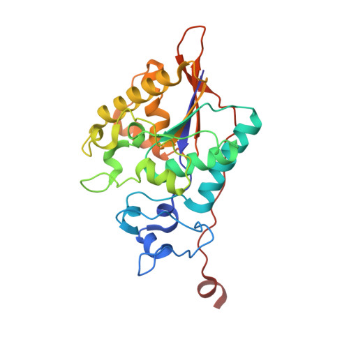
3MBK

The 1.35 A Structure of the Phosphatase Domain of the Suppressor of T Cell Receptor Signalling Protein in Complex with Sulphate


Experimental Data Snapshot

  • Method: X-RAY DIFFRACTION
  • Resolution: 1.35 Å
  • R-Value Free: 0.191 
  • R-Value Work: 0.162 
  • R-Value Observed: 0.163 

wwPDB Validation   3D Report Full Report


This is version 1.3 of the entry. See complete history


Literature

The 1.35 A resolution structure of the phosphatase domain of the suppressor of T-cell receptor signaling protein in complex with sulfate.

Jakoncic, J.Sondgeroth, B.Carpino, N.Nassar, N.

(2010) Acta Crystallogr Sect F Struct Biol Cryst Commun 66: 643-647

  • DOI: https://doi.org/10.1107/S1744309110014259
  • Primary Citation of Related Structures:  
    3MBK

  • PubMed Abstract: 

    The suppressor of T-cell signaling (Sts) proteins are multidomain proteins that negatively regulate the signaling of membrane-bound receptors, including the T-cell receptor (TCR) and the epidermal growth-factor receptor (EGFR). They contain at their C-terminus a 2H-phosphatase homology (PGM) domain that is responsible for their protein tyrosine phosphatase activity. Here, the crystal structure of the phosphatase domain of Sts-1, Sts-1(PGM), was determined at pH 4.6. The asymmetric unit contains two independent molecules and each active site is occupied by a sulfate ion. Each sulfate is located at the phosphate-binding site and makes similar interactions with the catalytic residues. The structure suggests an explanation for the lower Michaelis-Menten constants at acidic pH.


  • Organizational Affiliation

    Brookhaven National Laboratory, National Synchrotron Light Source, Building 725, Upton, NY 11973, USA.


Macromolecules
Find similar proteins by:  (by identity cutoff)  |  3D Structure
Entity ID: 1
MoleculeChains Sequence LengthOrganismDetailsImage
Ubiquitin-associated and SH3 domain-containing protein B
A, B
264Mus musculusMutation(s): 0 
Gene Names: Ubash3b
UniProt
Find proteins for Q8BGG7 (Mus musculus)
Explore Q8BGG7 
Go to UniProtKB:  Q8BGG7
Entity Groups  
Sequence Clusters30% Identity50% Identity70% Identity90% Identity95% Identity100% Identity
UniProt GroupQ8BGG7
Sequence Annotations
Expand
  • Reference Sequence
Experimental Data & Validation

Experimental Data

  • Method: X-RAY DIFFRACTION
  • Resolution: 1.35 Å
  • R-Value Free: 0.191 
  • R-Value Work: 0.162 
  • R-Value Observed: 0.163 
  • Space Group: P 21 21 21
Unit Cell:
Length ( Å )Angle ( ˚ )
a = 62.966α = 90
b = 80.049β = 90
c = 105.448γ = 90
Software Package:
Software NamePurpose
DCS_X6Adata collection
MOLREPphasing
REFMACrefinement
HKL-2000data reduction
HKL-2000data scaling

Structure Validation

View Full Validation Report


Entry History 

Deposition Data

Revision History  (Full details and data files)

  • Version 1.0: 2010-06-23
    Type: Initial release
  • Version 1.1: 2011-07-13
    Changes: Version format compliance
  • Version 1.2: 2015-01-28
    Changes: Other
  • Version 1.3: 2023-09-06
    Changes: Data collection, Database references, Derived calculations, Refinement description