3LS3

Padron0.9-ON (fluorescent state)


Experimental Data Snapshot

  • Method: X-RAY DIFFRACTION
  • Resolution: 1.65 Å
  • R-Value Free: 0.200 
  • R-Value Work: 0.168 
  • R-Value Observed: 0.169 

wwPDB Validation   3D Report Full Report


This is version 2.0 of the entry. See complete history


Literature

Molecular basis of the light-driven switching of the photochromic fluorescent protein Padron.

Brakemann, T.Weber, G.Andresen, M.Groenhof, G.Stiel, A.C.Trowitzsch, S.Eggeling, C.Grubmuller, H.Hell, S.W.Wahl, M.C.Jakobs, S.

(2010) J Biol Chem 285: 14603-14609

  • DOI: https://doi.org/10.1074/jbc.M109.086314
  • Primary Citation of Related Structures:  
    3LS3, 3LSA

  • PubMed Abstract: 

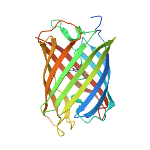
    Reversibly switchable fluorescent proteins can be repeatedly photoswitched between a fluorescent and a nonfluorescent state by irradiation with the light of two different wavelengths. The molecular basis of the switching process remains a controversial topic. Padron0.9 is a reversibly switchable fluorescent protein with "positive" switching characteristics, exhibiting excellent spectroscopic properties. Its chromophore is formed by the amino acids Cys-Tyr-Gly. We obtained high resolution x-ray structures of Padron0.9 in both the fluorescent and the nonfluorescent states and used the structural information for molecular dynamics simulations. We found that in Padron0.9 the chromophore undergoes a cis-trans isomerization upon photoswitching. The molecular dynamics simulations clarified the protonation states of the amino acid residues within the chromophore pocket that influence the protonation state of the chromophore. We conclude that a light driven cis-trans isomerization of the chromophore appears to be the fundamental switching mechanism in all photochromic fluorescent proteins known to date. Distinct absorption cross-sections for the switching wavelengths in the fluorescent and the nonfluorescent state are not essential for efficient photochromism in fluorescent proteins, although they may facilitate the switching process.


  • Organizational Affiliation

    Department of NanoBiophotonics, Max Planck Institute for Biophysical Chemistry, Am Fassberg 11, 37077 Göttingen, Germany.


Macromolecules
Find similar proteins by:  (by identity cutoff)  |  3D Structure
Entity ID: 1
MoleculeChains Sequence LengthOrganismDetailsImage
Padron0.9
A, B, C, D
235PectiniidaeMutation(s): 0 
Entity Groups  
Sequence Clusters30% Identity50% Identity70% Identity90% Identity95% Identity100% Identity
Sequence Annotations
Expand
  • Reference Sequence
Small Molecules
Ligands 3 Unique
IDChains Name / Formula / InChI Key2D Diagram3D Interactions
EPE
Query on EPE

Download Ideal Coordinates CCD File 
M [auth D]4-(2-HYDROXYETHYL)-1-PIPERAZINE ETHANESULFONIC ACID
C8 H18 N2 O4 S
JKMHFZQWWAIEOD-UHFFFAOYSA-N
SPD
Query on SPD

Download Ideal Coordinates CCD File 
G [auth A],
H [auth B],
I [auth B],
K [auth C],
L [auth C]
SPERMIDINE
C7 H19 N3
ATHGHQPFGPMSJY-UHFFFAOYSA-N
GOL
Query on GOL

Download Ideal Coordinates CCD File 
E [auth A],
F [auth A],
J [auth C],
N [auth D],
O [auth D]
GLYCEROL
C3 H8 O3
PEDCQBHIVMGVHV-UHFFFAOYSA-N
Modified Residues  1 Unique
IDChains TypeFormula2D DiagramParent
GYC
Query on GYC
A, B, C, D
L-PEPTIDE LINKINGC14 H15 N3 O4 SCYS, TYR, GLY
Experimental Data & Validation

Experimental Data

  • Method: X-RAY DIFFRACTION
  • Resolution: 1.65 Å
  • R-Value Free: 0.200 
  • R-Value Work: 0.168 
  • R-Value Observed: 0.169 
  • Space Group: P 21 21 21
Unit Cell:
Length ( Å )Angle ( ˚ )
a = 72.855α = 90
b = 103.806β = 90
c = 121.213γ = 90
Software Package:
Software NamePurpose
MAR345dtbdata collection
PHASERphasing
REFMACrefinement
HKL-2000data reduction
SCALEPACKdata scaling

Structure Validation

View Full Validation Report


Entry History 

Deposition Data

Revision History  (Full details and data files)

  • Version 1.0: 2010-03-16
    Type: Initial release
  • Version 1.1: 2011-07-13
    Changes: Advisory, Version format compliance
  • Version 1.2: 2023-09-06
    Changes: Data collection, Database references, Derived calculations, Refinement description
  • Version 2.0: 2023-11-15
    Changes: Advisory, Atomic model, Data collection, Derived calculations