3LHB

THE 2.7 ANGSTROM CRYSTAL STRUCTURE OF DEOXYGENATED HEMOGLOBIN FROM THE SEA LAMPREY (PETROMYZON MARINUS)


Experimental Data Snapshot

  • Method: X-RAY DIFFRACTION
  • Resolution: 2.70 Å
  • R-Value Free: 0.232 
  • R-Value Work: 0.195 
  • R-Value Observed: 0.195 

wwPDB Validation   3D Report Full Report


Ligand Structure Quality Assessment 


This is version 1.3 of the entry. See complete history


Literature

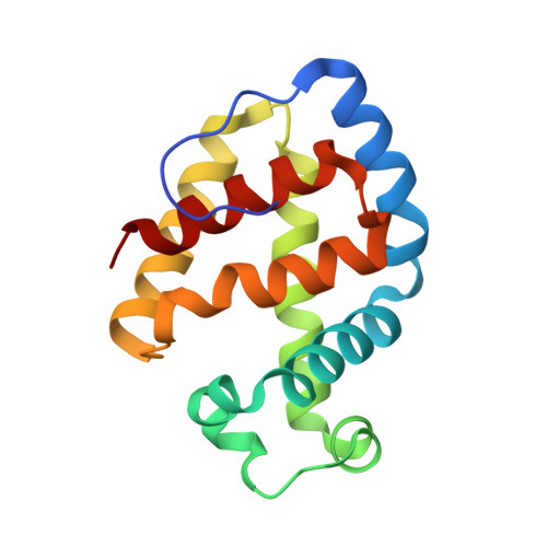
The 2.7 A crystal structure of deoxygenated hemoglobin from the sea lamprey (Petromyzon marinus): structural basis for a lowered oxygen affinity and Bohr effect.

Heaslet, H.A.Royer Jr., W.E.

(1999) Structure 7: 517-526

  • DOI: https://doi.org/10.1016/s0969-2126(99)80068-9
  • Primary Citation of Related Structures:  
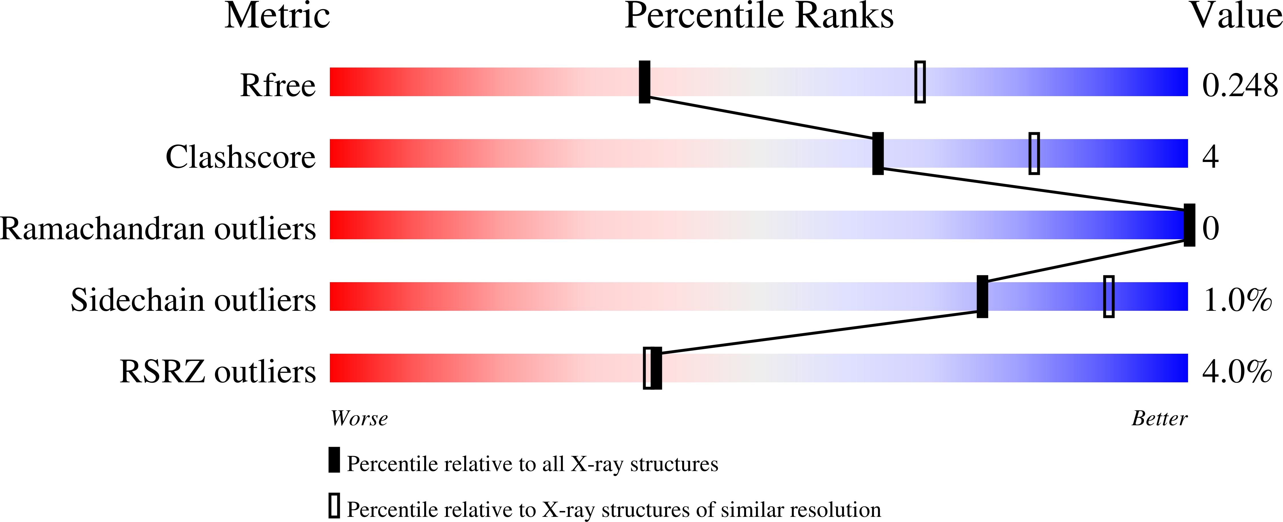
    3LHB

  • PubMed Abstract: 

    The hemoglobins of the sea lamprey are unusual in that cooperativity and sensitivity to pH arise from an equilibrium between a high-affinity monomer and a low-affinity oligomer. Although the crystal structure of the monomeric cyanide derivative has previously been determined, the manner by which oligomerization acts to lower the oxygen affinity and confer a strong Bohr effect has, until now, been speculative.


  • Organizational Affiliation

    Department of Biochemistry and Molecular Biology, University of Massachusetts Medical School, Worcester 01655, USA.


Macromolecules
Find similar proteins by:  (by identity cutoff)  |  3D Structure
Entity ID: 1
MoleculeChains Sequence LengthOrganismDetailsImage
PROTEIN (HEMOGLOBIN)
A, B, C, D, E
A, B, C, D, E, F, G, H, I, J, K, L
149Petromyzon marinusMutation(s): 0 
UniProt
Find proteins for P02208 (Petromyzon marinus)
Explore P02208 
Go to UniProtKB:  P02208
Entity Groups  
Sequence Clusters30% Identity50% Identity70% Identity90% Identity95% Identity100% Identity
UniProt GroupP02208
Sequence Annotations
Expand
  • Reference Sequence
Small Molecules
Ligands 1 Unique
IDChains Name / Formula / InChI Key2D Diagram3D Interactions
HEM
Query on HEM

Download Ideal Coordinates CCD File 
M [auth A]
N [auth B]
O [auth C]
P [auth D]
Q [auth E]
M [auth A],
N [auth B],
O [auth C],
P [auth D],
Q [auth E],
R [auth F],
S [auth G],
T [auth H],
U [auth I],
V [auth J],
W [auth K],
X [auth L]
PROTOPORPHYRIN IX CONTAINING FE
C34 H32 Fe N4 O4
KABFMIBPWCXCRK-RGGAHWMASA-L
Experimental Data & Validation

Experimental Data

  • Method: X-RAY DIFFRACTION
  • Resolution: 2.70 Å
  • R-Value Free: 0.232 
  • R-Value Work: 0.195 
  • R-Value Observed: 0.195 
  • Space Group: P 1 21 1
Unit Cell:
Length ( Å )Angle ( ˚ )
a = 59.63α = 90
b = 215.16β = 95.8
c = 75.39γ = 90
Software Package:
Software NamePurpose
DENZOdata reduction
SCALEPACKdata scaling
AMoREphasing
X-PLORrefinement

Structure Validation

View Full Validation Report


Ligand Structure Quality Assessment 


Entry History 

Deposition Data

Revision History  (Full details and data files)

  • Version 1.0: 1999-01-20
    Type: Initial release
  • Version 1.1: 2008-04-27
    Changes: Version format compliance
  • Version 1.2: 2011-07-13
    Changes: Version format compliance
  • Version 1.3: 2023-09-06
    Changes: Data collection, Database references, Derived calculations, Refinement description