Understanding blue-to-red conversion in monomeric fluorescent timers and hydrolytic degradation of their chromophores
Pletnev, S., Subach, F.V., Dauter, Z., Wlodawer, A., Verkhusha, V.V.(2010) J Am Chem Soc 132: 2243-2253
- PubMed: 20121102
- DOI: https://doi.org/10.1021/ja908418r
- Primary Citation of Related Structures:
3LF3, 3LF4 - PubMed Abstract:
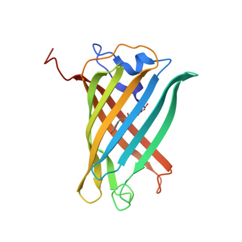
Fast-FT is a fluorescent timer (FT) engineered from DsRed-like fluorescent protein mCherry. Crystal structures of Fast-FT (chromophore Met66-Tyr67-Gly68) and its precursor with blocked blue-to-red conversion Blue102 (chromophore Leu66-Tyr67-Gly68) have been determined at the resolution of 1.15 A and 1.81 A, respectively. Structural data suggest that blue-to-red conversion, taking place in Fast-FT and in related FTs, is associated with the oxidation of Calpha2-Cbeta2 bond of Tyr67. Site directed mutagenesis revealed a crucial role of Arg70 and Tyr83 in the delayed oxidation of Calpha2-Cbeta2 bond, introducing the timing factor in maturation of the timer. Substitutions Ser217Ala and Ser217Cys in Fast-FT substantially slow down formation of an intermediate blue chromophore but do not affect much blue-to-red conversion, whereas mutations Arg70Lys or Trp83Leu, having little effect on the blue chromophore formation rate, markedly accelerates formation of the red chromophore. The chromophore of FTs adopts a cis-conformation stabilized by a hydrogen bond between its phenolate oxygen and the side chain hydroxyl of Ser146. In Blue102, a bulky side chain of Ile146 precludes the chromophore from adopting a "cis-like" conformation, blocking its blue-to-red conversion. Both Fast-FT and Blue102 structures revealed hydrolytic degradation of the chromophores. In Fast-FT, chromophore-forming Met66 residue is eliminated from the polypeptide chain, whereas Leu66 in Blue102 is cleaved out from the chromophore, decarboxylated and remains attached to the preceding Phe65. Hydrolysis of the chromophore competes with chromophore maturation and is driven by the same residues that participate in chromophore maturation.
Organizational Affiliation:
Synchrotron Radiation Research Section, Macromolecular Crystallography Laboratory, National Cancer Institute, Argonne, Illinois 60439, USA. pletnevs@mail.nih.gov