3L7I

Structure of the Wall Teichoic Acid Polymerase TagF


Experimental Data Snapshot

  • Method: X-RAY DIFFRACTION
  • Resolution: 2.70 Å
  • R-Value Free: 0.260 
  • R-Value Work: 0.210 
  • R-Value Observed: 0.213 

wwPDB Validation   3D Report Full Report


This is version 1.3 of the entry. See complete history


Literature

Structure of the bacterial teichoic acid polymerase TagF provides insights into membrane association and catalysis.

Lovering, A.L.Lin, L.Y.Sewell, E.W.Spreter, T.Brown, E.D.Strynadka, N.C.

(2010) Nat Struct Mol Biol 17: 582-589

  • DOI: https://doi.org/10.1038/nsmb.1819
  • Primary Citation of Related Structures:  
    3L7I, 3L7J, 3L7K, 3L7L, 3L7M

  • PubMed Abstract: 

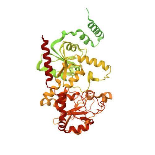
    Teichoic acid polymers are composed of polyol-phosphate units and form a major component of Gram-positive bacterial cell walls. These anionic compounds perform a multitude of important roles in bacteria and are synthesized by monotopic membrane proteins of the TagF polymerase family. We have determined the structure of Staphylococcus epidermidis TagF to 2.7-A resolution from a construct that includes both the membrane-targeting region and the glycerol-phosphate polymerase domains. TagF possesses a helical region for interaction with the lipid bilayer, placing the active site at a suitable distance for access to the membrane-bound substrate. Characterization of active-site residue variants and analysis of a CDP-glycerol substrate complex suggest a mechanism for polymer synthesis. With the importance of teichoic acid in Gram-positive physiology, this elucidation of the molecular details of TagF function provides a critical new target in the development of novel anti-infectives.


  • Organizational Affiliation

    Department of Biochemistry and Molecular Biology, Vancouver, Canada.


Macromolecules
Find similar proteins by:  (by identity cutoff)  |  3D Structure
Entity ID: 1
MoleculeChains Sequence LengthOrganismDetailsImage
Teichoic acid biosynthesis protein F
A, B, C, D
729Staphylococcus epidermidis RP62AMutation(s): 0 
Gene Names: SERP1960tagF
EC: 2.7.8.12
Membrane Entity: Yes 
UniProt
Find proteins for Q5HLM5 (Staphylococcus epidermidis (strain ATCC 35984 / RP62A))
Explore Q5HLM5 
Go to UniProtKB:  Q5HLM5
Entity Groups  
Sequence Clusters30% Identity50% Identity70% Identity90% Identity95% Identity100% Identity
UniProt GroupQ5HLM5
Sequence Annotations
Expand
  • Reference Sequence
Small Molecules
Ligands 3 Unique
IDChains Name / Formula / InChI Key2D Diagram3D Interactions
SO4
Query on SO4

Download Ideal Coordinates CCD File 
BA [auth C]
CA [auth C]
E [auth A]
F [auth A]
G [auth A]
BA [auth C],
CA [auth C],
E [auth A],
F [auth A],
G [auth A],
KA [auth D],
LA [auth D],
T [auth B]
SULFATE ION
O4 S
QAOWNCQODCNURD-UHFFFAOYSA-L
EDO
Query on EDO

Download Ideal Coordinates CCD File 
DA [auth C],
EA [auth C],
MA [auth D],
NA [auth D],
U [auth B]
1,2-ETHANEDIOL
C2 H6 O2
LYCAIKOWRPUZTN-UHFFFAOYSA-N
CL
Query on CL

Download Ideal Coordinates CCD File 
AA [auth B]
FA [auth C]
GA [auth C]
H [auth A]
HA [auth C]
AA [auth B],
FA [auth C],
GA [auth C],
H [auth A],
HA [auth C],
I [auth A],
IA [auth C],
J [auth A],
JA [auth C],
K [auth A],
L [auth A],
M [auth A],
N [auth A],
O [auth A],
OA [auth D],
P [auth A],
PA [auth D],
Q [auth A],
QA [auth D],
R [auth A],
S [auth A],
V [auth B],
W [auth B],
X [auth B],
Y [auth B],
Z [auth B]
CHLORIDE ION
Cl
VEXZGXHMUGYJMC-UHFFFAOYSA-M
Experimental Data & Validation

Experimental Data

  • Method: X-RAY DIFFRACTION
  • Resolution: 2.70 Å
  • R-Value Free: 0.260 
  • R-Value Work: 0.210 
  • R-Value Observed: 0.213 
  • Space Group: P 41 21 2
Unit Cell:
Length ( Å )Angle ( ˚ )
a = 223.739α = 90
b = 223.739β = 90
c = 100.691γ = 90
Software Package:
Software NamePurpose
MAR345dtbdata collection
SHELXSphasing
PHENIXrefinement
MOSFLMdata reduction
SCALAdata scaling

Structure Validation

View Full Validation Report


Entry History 

Deposition Data

Revision History  (Full details and data files)

  • Version 1.0: 2010-04-28
    Type: Initial release
  • Version 1.1: 2011-07-13
    Changes: Version format compliance
  • Version 1.2: 2014-11-12
    Changes: Experimental preparation
  • Version 1.3: 2024-02-21
    Changes: Data collection, Database references, Derived calculations