3L3P

Crystal structure of the C-terminal domain of Shigella type III effector IpaH9.8, with a novel domain swap


Experimental Data Snapshot

  • Method: X-RAY DIFFRACTION
  • Resolution: 3.20 Å
  • R-Value Free: 0.311 
  • R-Value Work: 0.289 
  • R-Value Observed: 0.290 

wwPDB Validation   3D Report Full Report


This is version 1.2 of the entry. See complete history


Literature

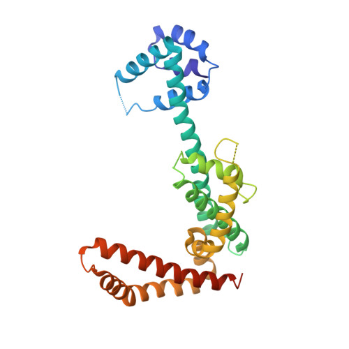
A disulfide driven domain swap switches off the activity of Shigella IpaH9.8 E3 ligase

Seyedarabi, A.Sullivan, J.A.Sasakawa, C.Pickersgill, R.W.

(2010) FEBS Lett 584: 4163-4168

  • DOI: https://doi.org/10.1016/j.febslet.2010.09.006
  • Primary Citation of Related Structures:  
    3L3P

  • PubMed Abstract: 

    We show that the monomeric form of Shigella IpaH9.8 E3 ligase catalyses the ubiquitination of human U2AF35 in vitro, providing a molecular mechanism for the observed in vivo effect. We further discover that under non-reducing conditions IpaH9.8 undergoes a domain swap driven by the formation of a disulfide bridge involving the catalytic cysteine and that this dimer is unable to catalyse the ubiquitination of U2AF35. The crystal structure of the domain-swapped dimer is presented. The redox inactivation of IpaH9.8 could be a mechanism of regulating the activity of the IpaH9.8 E3 ligase in response to cell damage so that the host cell in which the bacteria resides is maintained in a benign state suitable for bacterial survival.


  • Organizational Affiliation

    School of Biological and Chemical Sciences, Queen Mary University of London, London, United Kingdom. a.seyedarabi@qmul.ac.uk


Macromolecules
Find similar proteins by:  (by identity cutoff)  |  3D Structure
Entity ID: 1
MoleculeChains Sequence LengthOrganismDetailsImage
IpaH9.8293Shigella flexneriMutation(s): 0 
Gene Names: ipaH9.8
EC: 6.3.2.19
UniProt
Find proteins for Q8VSC3 (Shigella flexneri)
Explore Q8VSC3 
Go to UniProtKB:  Q8VSC3
Entity Groups  
Sequence Clusters30% Identity50% Identity70% Identity90% Identity95% Identity100% Identity
UniProt GroupQ8VSC3
Sequence Annotations
Expand
  • Reference Sequence
Experimental Data & Validation

Experimental Data

  • Method: X-RAY DIFFRACTION
  • Resolution: 3.20 Å
  • R-Value Free: 0.311 
  • R-Value Work: 0.289 
  • R-Value Observed: 0.290 
  • Space Group: P 62 2 2
Unit Cell:
Length ( Å )Angle ( ˚ )
a = 92.659α = 90
b = 92.659β = 90
c = 135.612γ = 120
Software Package:
Software NamePurpose
MOLREPphasing
REFMACrefinement
MOSFLMdata reduction
SCALAdata scaling

Structure Validation

View Full Validation Report


Entry History 

Deposition Data

Revision History  (Full details and data files)

  • Version 1.0: 2010-09-08
    Type: Initial release
  • Version 1.1: 2011-07-13
    Changes: Version format compliance
  • Version 1.2: 2023-11-01
    Changes: Data collection, Database references, Refinement description