3L3D

Crystal structure of HLA-B*4402 in complex with the F3A mutant of a self-peptide derived from DPA*0201


Experimental Data Snapshot

  • Method: X-RAY DIFFRACTION
  • Resolution: 1.80 Å
  • R-Value Free: 0.231 
  • R-Value Work: 0.188 
  • R-Value Observed: 0.189 

wwPDB Validation   3D Report Full Report


This is version 1.4 of the entry. See complete history


Literature

Constraints within major histocompatibility complex class I restricted peptides: presentation and consequences for T-cell recognition

Theodossis, A.Guillonneau, C.Welland, A.Ely, L.K.Clements, C.S.Williamson, N.A.Webb, A.I.Wilce, J.A.Mulder, R.J.Dunstone, M.A.Doherty, P.C.McCluskey, J.Purcell, A.W.Turner, S.J.Rossjohn, J.

(2010) Proc Natl Acad Sci U S A 107: 5534-5539

  • DOI: https://doi.org/10.1073/pnas.1000032107
  • Primary Citation of Related Structures:  
    3L3D, 3L3G, 3L3H, 3L3I, 3L3J, 3L3K

  • PubMed Abstract: 

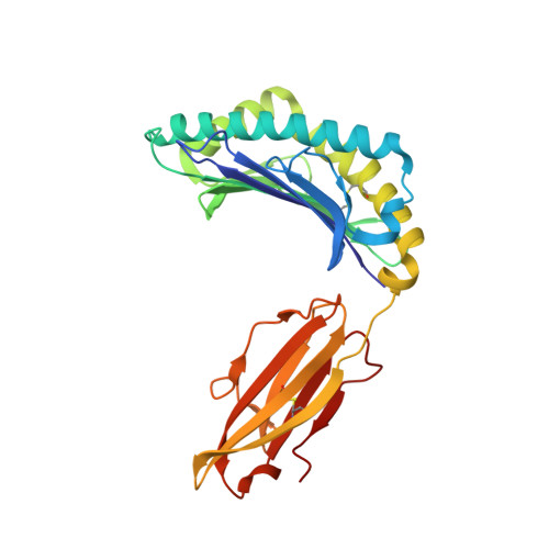
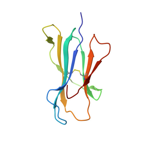
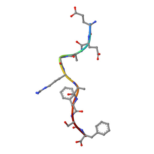
    Residues within processed protein fragments bound to major histocompatibility complex class I (MHC-I) glycoproteins have been considered to function as a series of "independent pegs" that either anchor the peptide (p) to the MHC-I and/or interact with the spectrum of alphabeta-T-cell receptors (TCRs) specific for the pMHC-I epitope in question. Mining of the extensive pMHC-I structural database established that many self- and viral peptides show extensive and direct interresidue interactions, an unexpected finding that has led us to the idea of "constrained" peptides. Mutational analysis of two constrained peptides (the HLA B44 restricted self-peptide (B44DPalpha-EEFGRAFSF) and an H2-D(b) restricted influenza peptide (D(b)PA, SSLENFRAYV) demonstrated that the conformation of the prominently exposed arginine in both peptides was governed by interactions with MHC-I-orientated flanking residues from the peptide itself. Using reverse genetics in a murine influenza model, we revealed that mutation of an MHC-I-orientated residue (SSLENFRAYV --> SSLENARAYV) within the constrained PA peptide resulted in a diminished cytotoxic T lymphocyte (CTL) response and the recruitment of a limited pMHC-I specific TCR repertoire. Interactions between individual peptide positions can thus impose fine control on the conformation of pMHC-I epitopes, whereas the perturbation of such constraints can lead to a previously unappreciated mechanism of viral escape.


  • Organizational Affiliation

    The Protein Crystallography Unit, Department of Biochemistry and Molecular Biology, School of Biomedical Sciences, Monash University, Victoria 3800, Australia.


Macromolecules
Find similar proteins by:  (by identity cutoff)  |  3D Structure
Entity ID: 1
MoleculeChains Sequence LengthOrganismDetailsImage
HLA class I histocompatibility antigen, B-44 alpha chain276Homo sapiensMutation(s): 0 
Gene Names: HLA-BHLAB
UniProt & NIH Common Fund Data Resources
Find proteins for P01889 (Homo sapiens)
Explore P01889 
Go to UniProtKB:  P01889
PHAROS:  P01889
GTEx:  ENSG00000234745 
Entity Groups  
Sequence Clusters30% Identity50% Identity70% Identity90% Identity95% Identity100% Identity
UniProt GroupP01889
Sequence Annotations
Expand
  • Reference Sequence
Find similar proteins by:  (by identity cutoff)  |  3D Structure
Entity ID: 2
MoleculeChains Sequence LengthOrganismDetailsImage
Beta-2-microglobulin99Homo sapiensMutation(s): 0 
Gene Names: B2MCDABP0092HDCMA22P
UniProt & NIH Common Fund Data Resources
Find proteins for P61769 (Homo sapiens)
Explore P61769 
Go to UniProtKB:  P61769
PHAROS:  P61769
GTEx:  ENSG00000166710 
Entity Groups  
Sequence Clusters30% Identity50% Identity70% Identity90% Identity95% Identity100% Identity
UniProt GroupP61769
Sequence Annotations
Expand
  • Reference Sequence

Find similar proteins by:  Sequence   |   3D Structure  

Entity ID: 3
MoleculeChains Sequence LengthOrganismDetailsImage
peptide from HLA-DPA1 protein9N/AMutation(s): 1 
UniProt
Find proteins for Q95HB9 (Homo sapiens)
Explore Q95HB9 
Go to UniProtKB:  Q95HB9
Entity Groups  
UniProt GroupQ95HB9
Sequence Annotations
Expand
  • Reference Sequence
Small Molecules
Experimental Data & Validation

Experimental Data

  • Method: X-RAY DIFFRACTION
  • Resolution: 1.80 Å
  • R-Value Free: 0.231 
  • R-Value Work: 0.188 
  • R-Value Observed: 0.189 
  • Space Group: P 21 21 21
Unit Cell:
Length ( Å )Angle ( ˚ )
a = 50.687α = 90
b = 82.019β = 90
c = 110.44γ = 90
Software Package:
Software NamePurpose
CrystalCleardata collection
AMoREphasing
REFMACrefinement
d*TREKdata reduction
d*TREKdata scaling

Structure Validation

View Full Validation Report


Entry History 

Deposition Data

Revision History  (Full details and data files)

  • Version 1.0: 2010-03-16
    Type: Initial release
  • Version 1.1: 2011-07-13
    Changes: Version format compliance
  • Version 1.2: 2014-02-26
    Changes: Database references
  • Version 1.3: 2021-11-10
    Changes: Database references, Derived calculations
  • Version 1.4: 2023-11-01
    Changes: Data collection, Refinement description