3L1O

Crystal structure of monoclonal antibody MN423 Fab fragment with free combining site, crystallized in the presence of zinc


Experimental Data Snapshot

  • Method: X-RAY DIFFRACTION
  • Resolution: 2.00 Å
  • R-Value Free: 0.249 
  • R-Value Work: 0.158 
  • R-Value Observed: 0.162 

wwPDB Validation   3D Report Full Report


This is version 1.2 of the entry. See complete history


Literature

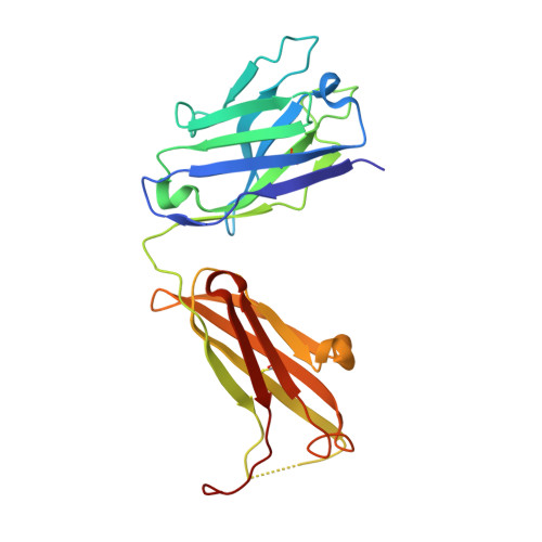
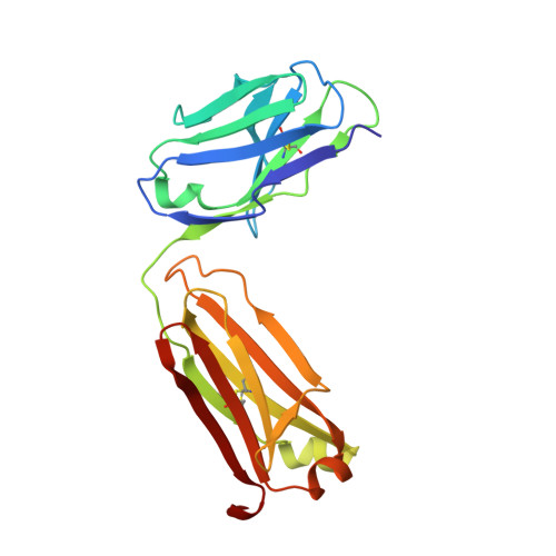
Monoclonal antibody MN423 as a stable mold facilitates structure determination of disordered tau protein

Skrabana, R.Dvorsky, R.Sevcik, J.Novak, M.

(2010) J Struct Biol 171: 74-81

  • DOI: https://doi.org/10.1016/j.jsb.2010.02.016
  • Primary Citation of Related Structures:  
    3L1O

  • PubMed Abstract: 

    Flexibility of intrinsically disordered tau protein is important for performing its functions. It is believed that alteration of the flexibility is instrumental to the assembly of tau protein into paired helical filaments (PHF) in tauopathies. Tau flexibility represents the main obstacle for structure determination of its conformation in physiology and/or pathology. We have alleviated this inherited difficulty by using specific monoclonal antibodies as tau protein surrogate binding partners. In this work we compare two "antibody mold structures": (1) X-ray structure of the free form of the Alzheimer's disease PHF core-specific antibody MN423 and (2) previously solved structure of the complex of MN423 with the PHF core C-terminal tau peptide. We found that MN423 combining site is in both structures identical. As a consequence, recombinant tau assumes in the complex a fold determined by the antibody combining site. Obtained results show that MN423 functions as a molecular mold for the PHF core segment, and opens the way for structure determination of other PHF core segments providing that other conformation-specific antibodies are available. Data from in silico docking of tau peptide into antibody mold, obtained in this study, show that biochemical data and computational approaches provide results comparable to X-ray crystallography.


  • Organizational Affiliation

    Institute of Neuroimmunology, Slovak Academy of Sciences, Dubravska cesta 9, 845 10 Bratislava, Slovakia.


Macromolecules
Find similar proteins by:  (by identity cutoff)  |  3D Structure
Entity ID: 1
MoleculeChains Sequence LengthOrganismDetailsImage
MONOCLONAL ANTIBODY FAB FRAGMENT MN423 H CHAINA [auth H]227Mus musculusMutation(s): 0 
Entity Groups  
Sequence Clusters30% Identity50% Identity70% Identity90% Identity95% Identity100% Identity
Sequence Annotations
Expand
  • Reference Sequence
Find similar proteins by:  (by identity cutoff)  |  3D Structure
Entity ID: 2
MoleculeChains Sequence LengthOrganismDetailsImage
MONOCLONAL ANTIBODY FAB FRAGMENT MN423 L CHAINB [auth L]214Mus musculusMutation(s): 0 
Entity Groups  
Sequence Clusters30% Identity50% Identity70% Identity90% Identity95% Identity100% Identity
Sequence Annotations
Expand
  • Reference Sequence
Experimental Data & Validation

Experimental Data

  • Method: X-RAY DIFFRACTION
  • Resolution: 2.00 Å
  • R-Value Free: 0.249 
  • R-Value Work: 0.158 
  • R-Value Observed: 0.162 
  • Space Group: P 1 21 1
Unit Cell:
Length ( Å )Angle ( ˚ )
a = 41.33α = 90
b = 75.426β = 92.86
c = 72.701γ = 90
Software Package:
Software NamePurpose
MAR345dtbdata collection
MOLREPphasing
REFMACrefinement
DENZOdata reduction
SCALEPACKdata scaling

Structure Validation

View Full Validation Report


Entry History 

Deposition Data

Revision History  (Full details and data files)

  • Version 1.0: 2010-03-16
    Type: Initial release
  • Version 1.1: 2011-07-13
    Changes: Advisory, Version format compliance
  • Version 1.2: 2023-11-01
    Changes: Data collection, Database references, Derived calculations, Refinement description