3L0I

Complex structure of SidM/DrrA with the wild type Rab1


Experimental Data Snapshot

  • Method: X-RAY DIFFRACTION
  • Resolution: 2.85 Å
  • R-Value Free: 0.266 
  • R-Value Work: 0.226 
  • R-Value Observed: 0.230 

wwPDB Validation   3D Report Full Report


This is version 1.2 of the entry. See complete history


Literature

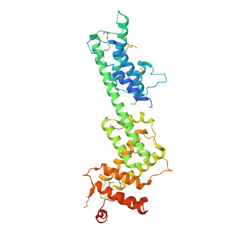
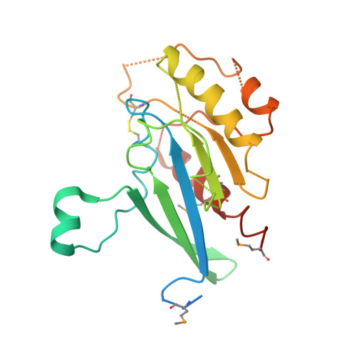
Structural mechanism of host Rab1 activation by the bifunctional Legionella type IV effector SidM/DrrA

Zhu, Y.Hu, L.Zhou, Y.Yao, Q.Liu, L.Shao, F.

(2010) Proc Natl Acad Sci U S A 107: 4699-4704

  • DOI: https://doi.org/10.1073/pnas.0914231107
  • Primary Citation of Related Structures:  
    3L0I, 3L0M

  • PubMed Abstract: 

    Bacterial pathogens deliver effector proteins with diverse biochemical activities into host cells, thereby modulating various host functions. Legionella pneumophila hijacks host vesicle trafficking to avoid phagosome-lysosome fusion, a mechanism that is dependent on the Legionella Dot/Icm type IV secretion system. SidM/DrrA, a Legionella type IV effector, is important for the interactions of Legionella-containing vacuoles with host endoplasmic reticulum-derived vesicles. SidM is the only known protein that catalyzes both the exchange of GDP for GTP and GDI displacement from small GTPase Rab1. We determined the crystal structures of SidM alone (residues 317-647) and SidM (residues 193-550) in complex with nucleotide-free WT Rab1. The SidM structure contains an N-terminal helical domain with a potential new function, a Rab1-activation domain, and a C-terminal phosphatidylinositol 4-phosphate-binding P4M domain. The Rab1-activation domain has extensive strong interactions mainly with Rab1 switch I and II regions that undergo substantial conformational changes on SidM binding. Mutations of switch-contacting residues in SidM attenuate both the nucleotide exchange and GDI displacement activities. Structural comparisons of Rab1 in the SidM complex with Rab1-GDP and Ypt1-GDP in the GDI complex identify key conformational changes that disrupt the nucleotide and GDI binding of Rab1. Further biochemical and structural analyses reveal a unique mechanism of coupled GDP release and GDI displacement likely triggered by the SidM-induced drastic displacement of switch I of Rab1.


  • Organizational Affiliation

    National Institute of Biological Sciences, Beijing 102206, China.


Macromolecules
Find similar proteins by:  (by identity cutoff)  |  3D Structure
Entity ID: 1
MoleculeChains Sequence LengthOrganismDetailsImage
DrrA
A, C
363Legionella pneumophilaMutation(s): 0 
Gene Names: sidM
UniProt
Find proteins for Q5ZSQ3 (Legionella pneumophila subsp. pneumophila (strain Philadelphia 1 / ATCC 33152 / DSM 7513))
Explore Q5ZSQ3 
Go to UniProtKB:  Q5ZSQ3
Entity Groups  
Sequence Clusters30% Identity50% Identity70% Identity90% Identity95% Identity100% Identity
UniProt GroupQ5ZSQ3
Sequence Annotations
Expand
  • Reference Sequence
Find similar proteins by:  (by identity cutoff)  |  3D Structure
Entity ID: 2
MoleculeChains Sequence LengthOrganismDetailsImage
Ras-related protein Rab-1A
B, D
199Homo sapiensMutation(s): 0 
Gene Names: RAB1A
UniProt & NIH Common Fund Data Resources
Find proteins for P62820 (Homo sapiens)
Explore P62820 
Go to UniProtKB:  P62820
PHAROS:  P62820
GTEx:  ENSG00000138069 
Entity Groups  
Sequence Clusters30% Identity50% Identity70% Identity90% Identity95% Identity100% Identity
UniProt GroupP62820
Sequence Annotations
Expand
  • Reference Sequence
Experimental Data & Validation

Experimental Data

  • Method: X-RAY DIFFRACTION
  • Resolution: 2.85 Å
  • R-Value Free: 0.266 
  • R-Value Work: 0.226 
  • R-Value Observed: 0.230 
  • Space Group: P 1 21 1
Unit Cell:
Length ( Å )Angle ( ˚ )
a = 63.808α = 90
b = 71.747β = 93.62
c = 146.911γ = 90
Software Package:
Software NamePurpose
DENZOdata reduction
SCALEPACKdata scaling
REFMACrefinement
PDB_EXTRACTdata extraction
ADSCdata collection
HKL-2000data reduction
SOLVEphasing

Structure Validation

View Full Validation Report


Entry History 

Deposition Data

Revision History  (Full details and data files)

  • Version 1.0: 2009-12-22
    Type: Initial release
  • Version 1.1: 2011-07-13
    Changes: Advisory, Version format compliance
  • Version 1.2: 2017-11-01
    Changes: Refinement description