3KWG

X-ray structure of NS1 effector domain W187A mutant


Experimental Data Snapshot

  • Method: X-RAY DIFFRACTION
  • Resolution: 2.21 Å
  • R-Value Free: 0.233 
  • R-Value Work: 0.187 
  • R-Value Observed: 0.189 

wwPDB Validation   3D Report Full Report


This is version 1.3 of the entry. See complete history


Literature

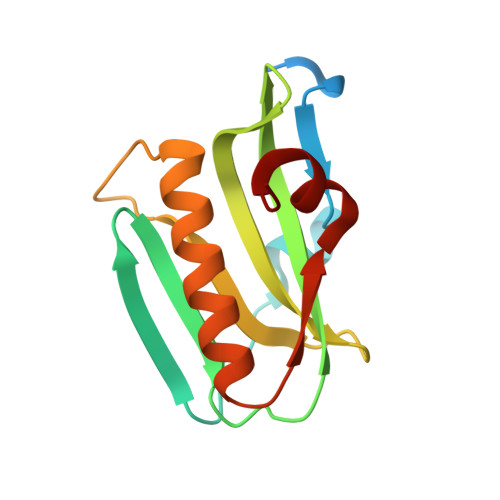
X-ray structures of NS1 effector domain mutants.

Xia, S.Robertus, J.D.

(2010) Arch Biochem Biophys 494: 198-204

  • DOI: https://doi.org/10.1016/j.abb.2009.12.008
  • Primary Citation of Related Structures:  
    3KWG, 3KWI

  • PubMed Abstract: 

    The influenza A virus nonstructural protein NS1 is a multifunctional dimeric protein that acts as a potent inhibitor of the host cellular antiviral state. The C-terminal effector domain of NS1 binds host proteins, including CPSF30, and is a target for the development of new antiviral drugs. Here we present crystallographic structures of two mutant effector domains, W187Y and W187A, of influenza A/Udorn/72 virus. Unlike wild-type, the mutants behave exclusively as monomers in solution based on gel filtration data and light scattering. The W187Y mutant is able to bind CPSF30 with a binding affinity close to the wild-type protein; that is, it retains a receptor site for aromatic ligands nearly identical to the wild-type. Therefore, this monomeric mutant protein could serve as a drug target for a high throughput inhibitor screening assays, since its binding pocket is unoccupied in solution and potentially more accessible to small molecule ligands.


  • Organizational Affiliation

    Institute for Cellular and Molecular Biology, Department of Chemistry and Biochemistry, University of Texas, 1 University Station A5300, Austin, TX 78712, USA.


Macromolecules
Find similar proteins by:  (by identity cutoff)  |  3D Structure
Entity ID: 1
MoleculeChains Sequence LengthOrganismDetailsImage
Non-structural protein 1
A, B
141Influenza A virus (A/Udorn/307/1972(H3N2))Mutation(s): 1 
Gene Names: NS
UniProt
Find proteins for P03495 (Influenza A virus (strain A/Udorn/307/1972 H3N2))
Explore P03495 
Go to UniProtKB:  P03495
Entity Groups  
Sequence Clusters30% Identity50% Identity70% Identity90% Identity95% Identity100% Identity
UniProt GroupP03495
Sequence Annotations
Expand
  • Reference Sequence
Experimental Data & Validation

Experimental Data

  • Method: X-RAY DIFFRACTION
  • Resolution: 2.21 Å
  • R-Value Free: 0.233 
  • R-Value Work: 0.187 
  • R-Value Observed: 0.189 
  • Space Group: P 21 21 21
Unit Cell:
Length ( Å )Angle ( ˚ )
a = 47.784α = 90
b = 48.193β = 90
c = 154.687γ = 90
Software Package:
Software NamePurpose
MAR345dtbdata collection
AMoREphasing
REFMACrefinement
HKL-2000data reduction
HKL-2000data scaling

Structure Validation

View Full Validation Report


Entry History 

Deposition Data

Revision History  (Full details and data files)

  • Version 1.0: 2010-02-02
    Type: Initial release
  • Version 1.1: 2011-07-13
    Changes: Version format compliance
  • Version 1.2: 2021-10-13
    Changes: Database references
  • Version 1.3: 2023-09-06
    Changes: Data collection, Refinement description