3KT4

Crystal structure of Tpa1 from Saccharomyces cerevisiae, a component of the messenger ribonucleoprotein complex


Experimental Data Snapshot

  • Method: X-RAY DIFFRACTION
  • Resolution: 2.73 Å
  • R-Value Free: 0.247 
  • R-Value Work: 0.190 
  • R-Value Observed: 0.193 

wwPDB Validation   3D Report Full Report


This is version 1.3 of the entry. See complete history


Literature

Crystal structure of Tpa1 from Saccharomyces cerevisiae, a component of the messenger ribonucleoprotein complex

Kim, H.S.Kim, H.L.Kim, K.H.Kim, D.J.Lee, S.J.Yoon, J.Y.Yoon, H.J.Lee, H.Y.Park, S.B.Kim, S.-J.Lee, J.Y.Suh, S.W.

(2010) Nucleic Acids Res 38: 2099-2110

  • DOI: https://doi.org/10.1093/nar/gkp1151
  • Primary Citation of Related Structures:  
    3KT1, 3KT4, 3KT7

  • PubMed Abstract: 

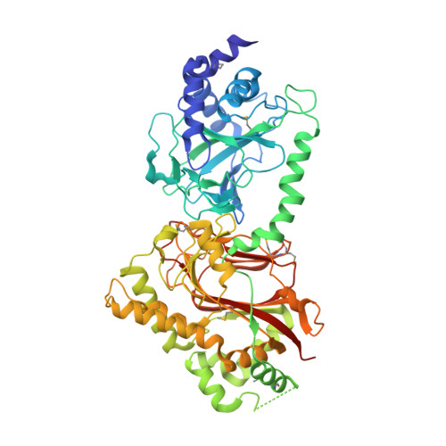
    Tpa1 (for termination and polyadenylation) from Saccharomyces cerevisiae is a component of a messenger ribonucleoprotein (mRNP) complex at the 3' untranslated region of mRNAs. It comprises an N-terminal Fe(II)- and 2-oxoglutarate (2OG) dependent dioxygenase domain and a C-terminal domain. The N-terminal dioxygenase domain of a homologous Ofd1 protein from Schizosaccharomyces pombe was proposed to serve as an oxygen sensor that regulates the activity of the C-terminal degradation domain. Members of the Tpa1 family are also present in higher eukaryotes including humans. Here we report the crystal structure of S. cerevisiae Tpa1 as a representative member of the Tpa1 family. Structures have been determined as a binary complex with Fe(III) and as a ternary complex with Fe(III) and 2OG. The structures reveal that both domains of Tpa1 have the double-stranded beta-helix fold and are similar to prolyl 4-hydroxylases. However, the binding of Fe(III) and 2OG is observed in the N-terminal domain only. We also show that Tpa1 binds to poly(rA), suggesting its direct interaction with mRNA in the mRNP complex. The structural and functional data reported in this study support a role of the Tpa1 family as a hydroxylase in the mRNP complex and as an oxygen sensor.


  • Organizational Affiliation

    Department of Chemistry, College of Natural Sciences, Seoul National University, Seoul 151-742, Korea.


Macromolecules
Find similar proteins by:  (by identity cutoff)  |  3D Structure
Entity ID: 1
MoleculeChains Sequence LengthOrganismDetailsImage
PKHD-type hydroxylase TPA1633Saccharomyces cerevisiaeMutation(s): 0 
Gene Names: TPA1YER049W
EC: 1.14.11
UniProt
Find proteins for P40032 (Saccharomyces cerevisiae (strain ATCC 204508 / S288c))
Explore P40032 
Go to UniProtKB:  P40032
Entity Groups  
Sequence Clusters30% Identity50% Identity70% Identity90% Identity95% Identity100% Identity
UniProt GroupP40032
Sequence Annotations
Expand
  • Reference Sequence
Small Molecules
Ligands 1 Unique
IDChains Name / Formula / InChI Key2D Diagram3D Interactions
FE
Query on FE

Download Ideal Coordinates CCD File 
B [auth A]FE (III) ION
Fe
VTLYFUHAOXGGBS-UHFFFAOYSA-N
Modified Residues  1 Unique
IDChains TypeFormula2D DiagramParent
MSE
Query on MSE
A
L-PEPTIDE LINKINGC5 H11 N O2 SeMET
Experimental Data & Validation

Experimental Data

  • Method: X-RAY DIFFRACTION
  • Resolution: 2.73 Å
  • R-Value Free: 0.247 
  • R-Value Work: 0.190 
  • R-Value Observed: 0.193 
  • Space Group: P 3 2 1
Unit Cell:
Length ( Å )Angle ( ˚ )
a = 136.293α = 90
b = 136.293β = 90
c = 83.282γ = 120
Software Package:
Software NamePurpose
DENZOdata reduction
SCALEPACKdata scaling
REFMACrefinement
PDB_EXTRACTdata extraction
HKL-2000data collection
HKL-2000data reduction
SOLVEphasing

Structure Validation

View Full Validation Report


Entry History 

Deposition Data

Revision History  (Full details and data files)

  • Version 1.0: 2010-01-19
    Type: Initial release
  • Version 1.1: 2011-07-13
    Changes: Version format compliance
  • Version 1.2: 2014-02-26
    Changes: Database references
  • Version 1.3: 2017-11-01
    Changes: Refinement description