3KS0

Crystal structure of the heme domain of flavocytochrome b2 in complex with Fab B2B4


Experimental Data Snapshot

  • Method: X-RAY DIFFRACTION
  • Resolution: 2.70 Å
  • R-Value Free: 0.289 
  • R-Value Work: 0.214 
  • R-Value Observed: 0.218 

wwPDB Validation   3D Report Full Report


Ligand Structure Quality Assessment 


This is version 1.2 of the entry. See complete history


Literature

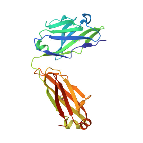
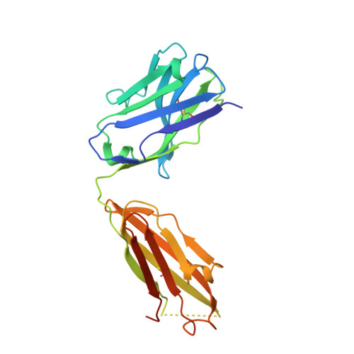
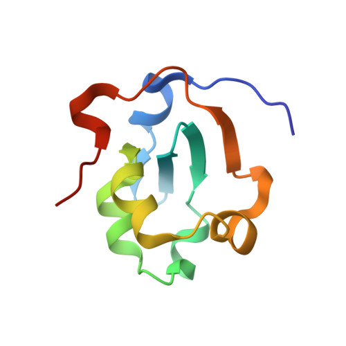
Structural evidence for the functional importance of the heme domain mobility in flavocytochrome b2.

Diep Le, K.H.Lederer, F.Golinelli-Pimpaneau, B.

(2010) J Mol Biol 400: 518-530

  • DOI: https://doi.org/10.1016/j.jmb.2010.05.035
  • Primary Citation of Related Structures:  
    3KS0

  • PubMed Abstract: 

    Yeast flavocytochrome b(2) (Fcb2) is an L-lactate:cytochrome c oxidoreductase in the mitochondrial intermembrane space participating in cellular respiration. Each enzyme subunit consists of a cytochrome b(5)-like heme domain and a flavodehydrogenase (FDH) domain. In the Fcb2 crystal structure, the heme domain is mobile relative to the tetrameric FDH core in one out of two subunits. The monoclonal antibody B2B4, elicited against the holoenzyme, recognizes only the native heme domain in the holoenzyme. When bound, it suppresses the intramolecular electron transfer from flavin to heme b(2), hence cytochrome c reduction. We report here the crystal structure of the heme domain in complex with the Fab at 2.7 A resolution. The Fab epitope on the heme domain includes the two exposed propionate groups of the heme, which are hidden in the interface between the domains in the complete subunit. The structure discloses an unexpected plasticity of Fcb2 in the neighborhood of the heme cavity, in which the heme has rotated. The epitope overlaps with the docking area of the FDH domain onto the heme domain, indicating that the antibody displaces the heme domain in a movement of large amplitude. We suggest that the binding sites on the heme domain of cytochrome c and of the FDH domain also overlap and therefore that cytochrome c binding also requires the heme domain to move away from the FDH domain, so as to allow electron transfer between the two hemes. Based on this hypothesis, we propose a possible model of the Fcb2.cytochrome c complex. Interestingly, this model shares similarity with that of the cytochrome b(5) x cytochrome c complex, in which cytochrome c binds to the surface around the exposed heme edge of cytochrome b(5). The present results therefore support the idea that the heme domain mobility is an inherent component of the Fcb2 functioning.


  • Organizational Affiliation

    Laboratoire d'Enzymologie et Biochimie Structurales, CNRS, 1 avenue de la Terrasse, 91198 Gif-sur-Yvette, France.


Macromolecules
Find similar proteins by:  (by identity cutoff)  |  3D Structure
Entity ID: 1
MoleculeChains Sequence LengthOrganismDetailsImage
heme domain of flavocytochrome b2A [auth L],
D [auth J]
214Mus musculusMutation(s): 0 
Entity Groups  
Sequence Clusters30% Identity50% Identity70% Identity90% Identity95% Identity100% Identity
Sequence Annotations
Expand
  • Reference Sequence
Find similar proteins by:  (by identity cutoff)  |  3D Structure
Entity ID: 2
MoleculeChains Sequence LengthOrganismDetailsImage
Fragment Antigen Binding B2B4B [auth H],
E [auth K]
225Mus musculusMutation(s): 0 
Entity Groups  
Sequence Clusters30% Identity50% Identity70% Identity90% Identity95% Identity100% Identity
Sequence Annotations
Expand
  • Reference Sequence
Find similar proteins by:  (by identity cutoff)  |  3D Structure
Entity ID: 3
MoleculeChains Sequence LengthOrganismDetailsImage
Cytochrome b2, mitochondrialC [auth A],
F [auth B]
95Saccharomyces cerevisiaeMutation(s): 0 
Gene Names: CYB2YML054CYM9958.08C
EC: 1.1.2.3
UniProt
Find proteins for P00175 (Saccharomyces cerevisiae (strain ATCC 204508 / S288c))
Explore P00175 
Go to UniProtKB:  P00175
Entity Groups  
Sequence Clusters30% Identity50% Identity70% Identity90% Identity95% Identity100% Identity
UniProt GroupP00175
Sequence Annotations
Expand
  • Reference Sequence
Small Molecules
Ligands 1 Unique
IDChains Name / Formula / InChI Key2D Diagram3D Interactions
HEM
Query on HEM

Download Ideal Coordinates CCD File 
G [auth A],
H [auth B]
PROTOPORPHYRIN IX CONTAINING FE
C34 H32 Fe N4 O4
KABFMIBPWCXCRK-RGGAHWMASA-L
Experimental Data & Validation

Experimental Data

  • Method: X-RAY DIFFRACTION
  • Resolution: 2.70 Å
  • R-Value Free: 0.289 
  • R-Value Work: 0.214 
  • R-Value Observed: 0.218 
  • Space Group: P 1 21 1
Unit Cell:
Length ( Å )Angle ( ˚ )
a = 72.58α = 90
b = 83.68β = 96.8
c = 92.165γ = 90
Software Package:
Software NamePurpose
REFMACrefinement
SCALAdata scaling
PHASERphasing
CNSrefinement
PDB_EXTRACTdata extraction
MOSFLMdata reduction

Structure Validation

View Full Validation Report


Ligand Structure Quality Assessment 


Entry History 

Deposition Data

Revision History  (Full details and data files)

  • Version 1.0: 2010-05-26
    Type: Initial release
  • Version 1.1: 2011-07-13
    Changes: Version format compliance
  • Version 1.2: 2019-07-17
    Changes: Data collection, Refinement description