3KN5

Crystal structure of the C-terminal kinase domain of msk1 in complex with AMP-PNP


Experimental Data Snapshot

  • Method: X-RAY DIFFRACTION
  • Resolution: 2.40 Å
  • R-Value Free: 0.278 
  • R-Value Work: 0.208 
  • R-Value Observed: 0.211 

wwPDB Validation   3D Report Full Report


Ligand Structure Quality Assessment 


This is version 1.2 of the entry. See complete history


Literature

The crystal structure of the active form of the C-terminal kinase domain of mitogen- and stress-activated protein kinase 1.

Malakhova, M.D'Angelo, I.Kim, H.G.Kurinov, I.Bode, A.M.Dong, Z.

(2010) J Mol Biol 399: 41-52

  • DOI: https://doi.org/10.1016/j.jmb.2010.03.064
  • Primary Citation of Related Structures:  
    3KN5, 3KN6

  • PubMed Abstract: 

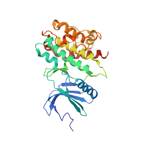
    Mitogen- and stress-activated protein kinase 1 (MSK1) is a growth-factor-stimulated serine/threonine kinase that is involved in gene transcription regulation and proinflammatory cytokine stimulation. MSK1 is a dual kinase possessing two nonidentical protein kinase domains in one polypeptide. We present the active conformation of the crystal structures of its C-terminal kinase domain in apo form and in complex with a nonhydrolyzable ATP analogue at 2.0 A and 2.5 A resolutions, respectively. Structural analysis revealed substantial differences in the contacts formed by the C-terminal helix, which is responsible for the inactivity of other autoinhibited kinases. In the C-terminal kinase domain of MSK1, the C-terminal alphaL-helix is located in the surface groove, but forms no hydrogen bonds with the substrate-binding loop or nearby helices, and does not interfere with the protein's autophosphorylation activity. Mutational analysis confirmed that the alphaL-helix is inherently nonautoinhibitory. Overexpression of the single C-terminal kinase domain in JB6 cells resulted in tumor-promoter-induced neoplastic transformation in a manner similar to that induced by the full-length MSK1 protein. The overall results suggest that the C-terminal kinase domain of MSK1 is regulated by a novel alphaL-helix-independent mechanism, suggesting that a diverse mechanism of autoinhibition and activation might be adopted by members of a closely related protein kinase family.


  • Organizational Affiliation

    The Hormel Institute University of Minnesota, 801 16th Avenue NE, Austin, MN 55912, USA.


Macromolecules
Find similar proteins by:  (by identity cutoff)  |  3D Structure
Entity ID: 1
MoleculeChains Sequence LengthOrganismDetailsImage
Ribosomal protein S6 kinase alpha-5
A, B
325Homo sapiensMutation(s): 0 
Gene Names: MSK1RPS6KA5
EC: 2.7.11.1
UniProt & NIH Common Fund Data Resources
Find proteins for O75582 (Homo sapiens)
Explore O75582 
Go to UniProtKB:  O75582
PHAROS:  O75582
GTEx:  ENSG00000100784 
Entity Groups  
Sequence Clusters30% Identity50% Identity70% Identity90% Identity95% Identity100% Identity
UniProt GroupO75582
Sequence Annotations
Expand
  • Reference Sequence
Small Molecules
Ligands 1 Unique
IDChains Name / Formula / InChI Key2D Diagram3D Interactions
ANP
Query on ANP

Download Ideal Coordinates CCD File 
C [auth A],
D [auth B]
PHOSPHOAMINOPHOSPHONIC ACID-ADENYLATE ESTER
C10 H17 N6 O12 P3
PVKSNHVPLWYQGJ-KQYNXXCUSA-N
Experimental Data & Validation

Experimental Data

  • Method: X-RAY DIFFRACTION
  • Resolution: 2.40 Å
  • R-Value Free: 0.278 
  • R-Value Work: 0.208 
  • R-Value Observed: 0.211 
  • Space Group: P 21 21 21
Unit Cell:
Length ( Å )Angle ( ˚ )
a = 51.561α = 90
b = 91.25β = 90
c = 135.46γ = 90
Software Package:
Software NamePurpose
PHASERphasing
PHENIXrefinement
HKL-2000data reduction
HKL-2000data scaling

Structure Validation

View Full Validation Report


Ligand Structure Quality Assessment 


Entry History 

Deposition Data

Revision History  (Full details and data files)

  • Version 1.0: 2010-04-21
    Type: Initial release
  • Version 1.1: 2011-07-13
    Changes: Version format compliance
  • Version 1.2: 2023-09-06
    Changes: Data collection, Database references, Derived calculations, Refinement description