3KKO

Crystal structure of M-Ras P40D/D41E/L51R in complex with GppNHp


Experimental Data Snapshot

  • Method: X-RAY DIFFRACTION
  • Resolution: 1.90 Å
  • R-Value Free: 0.222 
  • R-Value Work: 0.184 
  • R-Value Observed: 0.186 

wwPDB Validation   3D Report Full Report


Ligand Structure Quality Assessment 


This is version 1.3 of the entry. See complete history


Literature

Structural basis for conformational dynamics of GTP-bound Ras protein

Shima, F.Ijiri, Y.Muraoka, S.Liao, J.Ye, M.Araki, M.Matsumoto, K.Yamamoto, N.Sugimoto, T.Yoshikawa, Y.Kumasaka, T.Yamamoto, M.Tamura, A.Kataoka, T.

(2010) J Biol Chem 285: 22696-22705

  • DOI: https://doi.org/10.1074/jbc.M110.125161
  • Primary Citation of Related Structures:  
    3KKM, 3KKN, 3KKO, 3KKP, 3KKQ

  • PubMed Abstract: 

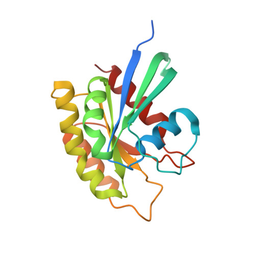
    Ras family small GTPases assume two interconverting conformations, "inactive" state 1 and "active" state 2, in their GTP-bound forms. Here, to clarify the mechanism of state transition, we have carried out x-ray crystal structure analyses of a series of mutant H-Ras and M-Ras in complex with guanosine 5'-(beta,gamma-imido)triphosphate (GppNHp), representing various intermediate states of the transition. Crystallization of H-RasT35S-GppNHp enables us to solve the first complete tertiary structure of H-Ras state 1 possessing two surface pockets unseen in the state 2 or H-Ras-GDP structure. Moreover, determination of the two distinct crystal structures of H-RasT35S-GppNHp, showing prominent polysterism in the switch I and switch II regions, reveals a pivotal role of the guanine nucleotide-mediated interaction between the two switch regions and its rearrangement by a nucleotide positional change in the state 2 to state 1 transition. Furthermore, the (31)P NMR spectra and crystal structures of the GppNHp-bound forms of M-Ras mutants, carrying various H-Ras-type amino acid substitutions, also reveal the existence of a surface pocket in state 1 and support a similar mechanism based on the nucleotide-mediated interaction and its rearrangement in the state 1 to state 2 transition. Intriguingly, the conformational changes accompanying the state transition mimic those that occurred upon GDP/GTP exchange, indicating a common mechanistic basis inherent in the high flexibility of the switch regions. Collectively, these results clarify the structural features distinguishing the two states and provide new insights into the molecular basis for the state transition of Ras protein.


  • Organizational Affiliation

    Division of Molecular Biology, Department of Biochemistry and Molecular Biology, Kobe University Graduate School of Medicine, 7-5-1 Kusunoki-cho, Chuo-ku, Kobe 650-0017, Japan.


Macromolecules
Find similar proteins by:  (by identity cutoff)  |  3D Structure
Entity ID: 1
MoleculeChains Sequence LengthOrganismDetailsImage
Ras-related protein M-RasA,
B,
C [auth P]
183Mus musculusMutation(s): 3 
UniProt & NIH Common Fund Data Resources
Find proteins for O08989 (Mus musculus)
Explore O08989 
Go to UniProtKB:  O08989
IMPC:  MGI:1100856
Entity Groups  
Sequence Clusters30% Identity50% Identity70% Identity90% Identity95% Identity100% Identity
UniProt GroupO08989
Sequence Annotations
Expand
  • Reference Sequence
Small Molecules
Ligands 3 Unique
IDChains Name / Formula / InChI Key2D Diagram3D Interactions
GNP
Query on GNP

Download Ideal Coordinates CCD File 
D [auth A],
G [auth B],
J [auth P]
PHOSPHOAMINOPHOSPHONIC ACID-GUANYLATE ESTER
C10 H17 N6 O13 P3
UQABYHGXWYXDTK-UUOKFMHZSA-N
SO4
Query on SO4

Download Ideal Coordinates CCD File 
F [auth A],
I [auth B],
L [auth P]
SULFATE ION
O4 S
QAOWNCQODCNURD-UHFFFAOYSA-L
MG
Query on MG

Download Ideal Coordinates CCD File 
E [auth A],
H [auth B],
K [auth P]
MAGNESIUM ION
Mg
JLVVSXFLKOJNIY-UHFFFAOYSA-N
Experimental Data & Validation

Experimental Data

  • Method: X-RAY DIFFRACTION
  • Resolution: 1.90 Å
  • R-Value Free: 0.222 
  • R-Value Work: 0.184 
  • R-Value Observed: 0.186 
  • Space Group: P 21 21 21
Unit Cell:
Length ( Å )Angle ( ˚ )
a = 78.196α = 90
b = 192.248β = 90
c = 43.235γ = 90
Software Package:
Software NamePurpose
BSSdata collection
MOLREPphasing
REFMACrefinement
MOSFLMdata reduction
SCALAdata scaling

Structure Validation

View Full Validation Report


Ligand Structure Quality Assessment 


Entry History 

Deposition Data

Revision History  (Full details and data files)

  • Version 1.0: 2010-06-16
    Type: Initial release
  • Version 1.1: 2011-07-13
    Changes: Version format compliance
  • Version 1.2: 2014-02-12
    Changes: Database references
  • Version 1.3: 2023-11-01
    Changes: Data collection, Database references, Derived calculations, Refinement description