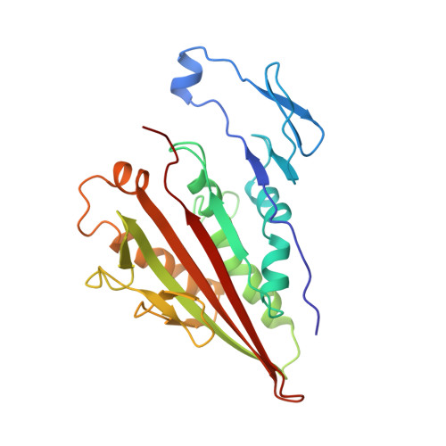
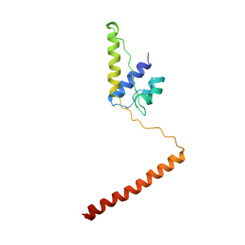
3KIN

KINESIN (DIMERIC) FROM RATTUS NORVEGICUS


Experimental Data Snapshot

  • Method: X-RAY DIFFRACTION
  • Resolution: 3.10 Å
  • R-Value Free: 0.362 
  • R-Value Observed: 0.289 

wwPDB Validation   3D Report Full Report


This is version 1.2 of the entry. See complete history


Literature

The crystal structure of dimeric kinesin and implications for microtubule-dependent motility.

Kozielski, F.Sack, S.Marx, A.Thormahlen, M.Schonbrunn, E.Biou, V.Thompson, A.Mandelkow, E.M.Mandelkow, E.

(1997) Cell 91: 985-994

  • DOI: https://doi.org/10.1016/s0092-8674(00)80489-4
  • Primary Citation of Related Structures:  
    3KIN

  • PubMed Abstract: 

    The dimeric form of the kinesin motor and neck domain from rat brain with bound ADP has been solved by X-ray crystallography. The two heads of the dimer are connected via a coiled-coil alpha-helical interaction of their necks. They are broadly similar to one another; differences are most apparent in the head-neck junction and in a moderate reorientation of the neck helices in order to adopt to the coiled-coil conformation. The heads show a rotational symmetry (approximately 120 degrees) about an axis close to that of the coiled-coil. This arrangement is unexpected since it is not compatible with the microtubule lattice. In this arrangement, the two heads of a kinesin dimer could not have equivalent interactions with microtubules.


  • Organizational Affiliation

    Max-Planck-Unit for Structural Molecular Biology, Hamburg, Germany.


Macromolecules
Find similar proteins by:  (by identity cutoff)  |  3D Structure
Entity ID: 1
MoleculeChains Sequence LengthOrganismDetailsImage
KINESIN HEAVY CHAIN
A, C
238Rattus norvegicusMutation(s): 0 
UniProt
Find proteins for P56536 (Rattus norvegicus)
Explore P56536 
Go to UniProtKB:  P56536
Entity Groups  
Sequence Clusters30% Identity50% Identity70% Identity90% Identity95% Identity100% Identity
UniProt GroupP56536
Sequence Annotations
Expand
  • Reference Sequence
Find similar proteins by:  (by identity cutoff)  |  3D Structure
Entity ID: 2
MoleculeChains Sequence LengthOrganismDetailsImage
KINESIN HEAVY CHAIN
B, D
117Rattus norvegicusMutation(s): 0 
UniProt
Find proteins for Q6QLM7 (Rattus norvegicus)
Explore Q6QLM7 
Go to UniProtKB:  Q6QLM7
Entity Groups  
Sequence Clusters30% Identity50% Identity70% Identity90% Identity95% Identity100% Identity
UniProt GroupQ6QLM7
Sequence Annotations
Expand
  • Reference Sequence
Small Molecules
Ligands 1 Unique
IDChains Name / Formula / InChI Key2D Diagram3D Interactions
ADP
Query on ADP

Download Ideal Coordinates CCD File 
E [auth A],
F [auth C]
ADENOSINE-5'-DIPHOSPHATE
C10 H15 N5 O10 P2
XTWYTFMLZFPYCI-KQYNXXCUSA-N
Experimental Data & Validation

Experimental Data

  • Method: X-RAY DIFFRACTION
  • Resolution: 3.10 Å
  • R-Value Free: 0.362 
  • R-Value Observed: 0.289 
  • Space Group: P 21 21 21
Unit Cell:
Length ( Å )Angle ( ˚ )
a = 72.15α = 90
b = 91.85β = 90
c = 141.68γ = 90
Software Package:
Software NamePurpose
PHASESphasing
REFMACrefinement
DENZOdata reduction
SCALEPACKdata scaling

Structure Validation

View Full Validation Report


Entry History 

Deposition Data

Revision History  (Full details and data files)

  • Version 1.0: 1998-10-14
    Type: Initial release
  • Version 1.1: 2008-03-25
    Changes: Version format compliance
  • Version 1.2: 2011-07-13
    Changes: Version format compliance