3JXT

Crystal structure of the third PDZ domain of SAP-102 in complex with a fluorogenic peptide-based ligand


Experimental Data Snapshot

  • Method: X-RAY DIFFRACTION
  • Resolution: 1.50 Å
  • R-Value Free: 0.214 
  • R-Value Work: 0.185 
  • R-Value Observed: 0.187 

wwPDB Validation   3D Report Full Report


This is version 1.5 of the entry. See complete history


Literature

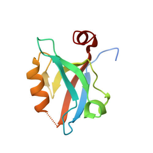
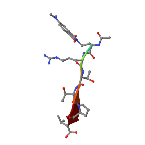
Caged mono- and divalent ligands for light-assisted disruption of PDZ domain-mediated interactions.

Sainlos, M.Iskenderian-Epps, W.S.Olivier, N.B.Choquet, D.Imperiali, B.

(2013) J Am Chem Soc 135: 4580-4583

  • DOI: https://doi.org/10.1021/ja309870q
  • Primary Citation of Related Structures:  
    3JXT

  • PubMed Abstract: 

    We report a general method for light-assisted control of interactions of PDZ domain binding motifs with their cognate domains by the incorporation of a photolabile caging group onto the essential C-terminal carboxylate binding determinant of the motif. The strategy was implemented and validated for both simple monovalent and biomimetic divalent ligands, which have recently been established as powerful tools for acute perturbation of native PDZ domain-dependent interactions in live cells.


  • Organizational Affiliation

    Departments of Chemistry and Biology, Massachusetts Institute of Technology, Cambridge, Massachusetts 02139, USA.


Macromolecules
Find similar proteins by:  (by identity cutoff)  |  3D Structure
Entity ID: 1
MoleculeChains Sequence LengthOrganismDetailsImage
Disks large homolog 3
A, B
104Rattus norvegicusMutation(s): 0 
Gene Names: Dlg3Dlgh3
UniProt
Find proteins for Q62936 (Rattus norvegicus)
Explore Q62936 
Go to UniProtKB:  Q62936
Entity Groups  
Sequence Clusters30% Identity50% Identity70% Identity90% Identity95% Identity100% Identity
UniProt GroupQ62936
Sequence Annotations
Expand
  • Reference Sequence

Find similar proteins by:  Sequence   |   3D Structure  

Entity ID: 2
MoleculeChains Sequence LengthOrganismDetailsImage
Voltage-dependent calcium channel gamma-2 subunit
C, D
7N/AMutation(s): 1 
UniProt & NIH Common Fund Data Resources
Find proteins for O88602 (Mus musculus)
Explore O88602 
Go to UniProtKB:  O88602
IMPC:  MGI:1316660
Entity Groups  
UniProt GroupO88602
Sequence Annotations
Expand
  • Reference Sequence
Small Molecules
Ligands 1 Unique
IDChains Name / Formula / InChI Key2D Diagram3D Interactions
ACT
Query on ACT

Download Ideal Coordinates CCD File 
E [auth A]ACETATE ION
C2 H3 O2
QTBSBXVTEAMEQO-UHFFFAOYSA-M
Experimental Data & Validation

Experimental Data

  • Method: X-RAY DIFFRACTION
  • Resolution: 1.50 Å
  • R-Value Free: 0.214 
  • R-Value Work: 0.185 
  • R-Value Observed: 0.187 
  • Space Group: P 21 21 21
Unit Cell:
Length ( Å )Angle ( ˚ )
a = 49.597α = 90
b = 54.215β = 90
c = 86.023γ = 90
Software Package:
Software NamePurpose
ADSCdata collection
MOLREPphasing
REFMACrefinement
HKL-2000data reduction
HKL-2000data scaling

Structure Validation

View Full Validation Report


Entry History 

Deposition Data

Revision History  (Full details and data files)

  • Version 1.0: 2010-09-29
    Type: Initial release
  • Version 1.1: 2011-07-13
    Changes: Version format compliance
  • Version 1.2: 2013-04-10
    Changes: Database references
  • Version 1.3: 2021-10-13
    Changes: Database references, Derived calculations
  • Version 1.4: 2023-09-06
    Changes: Data collection, Refinement description
  • Version 1.5: 2023-11-22
    Changes: Data collection