3JVG

Crystal Structure of chicken CD1-1


Experimental Data Snapshot

  • Method: X-RAY DIFFRACTION
  • Resolution: 2.20 Å
  • R-Value Free: 0.252 
  • R-Value Work: 0.216 
  • R-Value Observed: 0.218 

wwPDB Validation   3D Report Full Report


Ligand Structure Quality Assessment 


This is version 1.3 of the entry. See complete history


Literature

Structural basis for lipid-antigen recognition in avian immunity.

Dvir, H.Wang, J.Ly, N.Dascher, C.C.Zajonc, D.M.

(2010) J Immunol 184: 2504-2511

  • DOI: https://doi.org/10.4049/jimmunol.0903509
  • Primary Citation of Related Structures:  
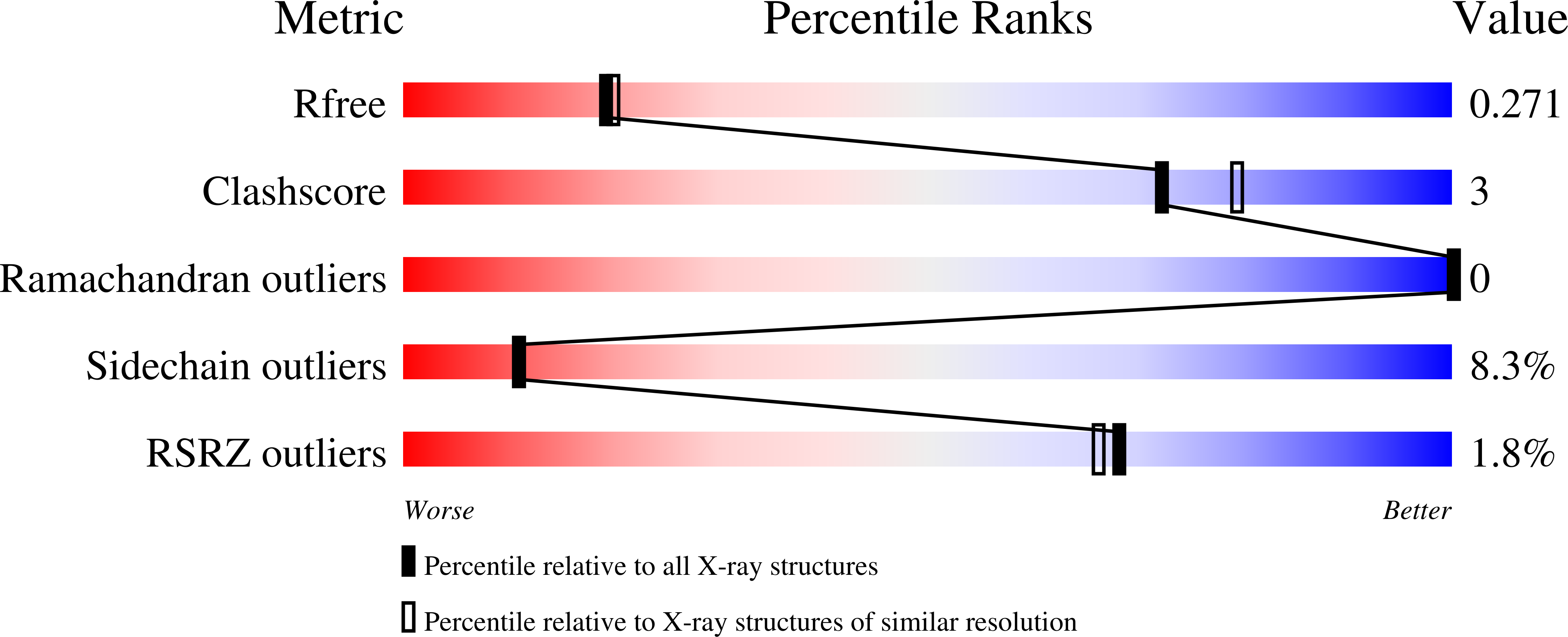
    3JVG

  • PubMed Abstract: 

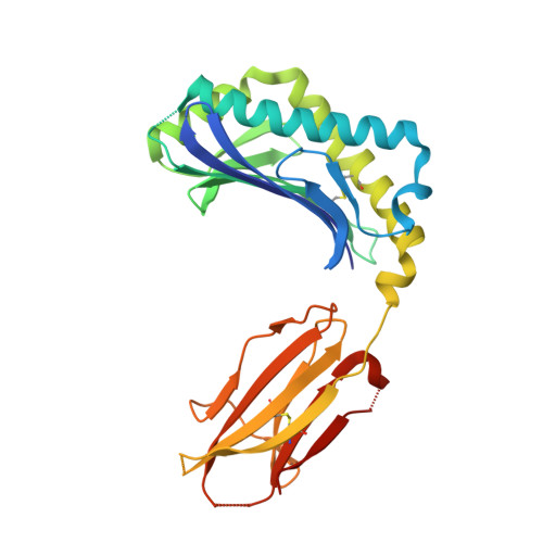
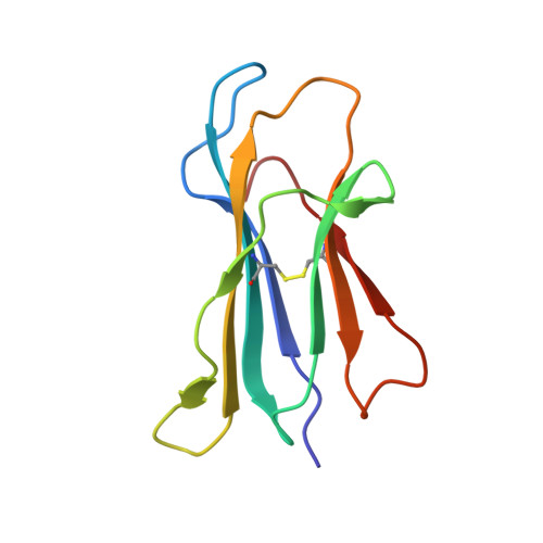
    CD1 proteins present self- and foreign lipid Ags to activate specific T cells in the mammalian immune system. These T cells play an important role in controlling autoimmune diseases, suppression of tumor growth, and host defense against invading pathogens. Humans use five CD1 isoforms, whereas only two exist in birds. Unlike mammals' CD1, the structure of chicken CD1-2 showed a primitive lipid-binding groove, suggesting that chicken may only recognize single-chain lipids. In contrast, the crystal structure of the second chicken CD1 isoform, chCD1-1, reported in this study at 2.2 A resolution, reveals an elaborated binding groove with a dual-pocket, dual-cleft architecture. The A' and F' deep pockets are separated from each other, but each is connected to a hydrophobic surface cleft, which may participate in lipid binding. The long endogenous ligand found inside the binding groove of chCD1-1, together with binding data on various glycolipids and mycolic acid, strongly suggest that the unique avian CD1 family could bind long dual- and possibly triacyl-chain lipids.


  • Organizational Affiliation

    Department of Cell Biology, La Jolla Institute for Allergy and Immunology, La Jolla CA 92037, USA.


Macromolecules
Find similar proteins by:  (by identity cutoff)  |  3D Structure
Entity ID: 1
MoleculeChains Sequence LengthOrganismDetailsImage
T-cell surface glycoprotein CD1A1 antigen
A, B
281Gallus gallusMutation(s): 0 
Gene Names: CD1A1chicken cd1.1
UniProt
Find proteins for A5HUM9 (Gallus gallus)
Explore A5HUM9 
Go to UniProtKB:  A5HUM9
Entity Groups  
Sequence Clusters30% Identity50% Identity70% Identity90% Identity95% Identity100% Identity
UniProt GroupA5HUM9
Sequence Annotations
Expand
  • Reference Sequence
Find similar proteins by:  (by identity cutoff)  |  3D Structure
Entity ID: 2
MoleculeChains Sequence LengthOrganismDetailsImage
Beta-2-microglobulin
C, D
98Gallus gallusMutation(s): 0 
Gene Names: B2Mbeta-2-microglobulin
UniProt
Find proteins for P21611 (Gallus gallus)
Explore P21611 
Go to UniProtKB:  P21611
Entity Groups  
Sequence Clusters30% Identity50% Identity70% Identity90% Identity95% Identity100% Identity
UniProt GroupP21611
Sequence Annotations
Expand
  • Reference Sequence
Small Molecules
Ligands 3 Unique
IDChains Name / Formula / InChI Key2D Diagram3D Interactions
NAG
Query on NAG

Download Ideal Coordinates CCD File 
H [auth A]
I [auth A]
J [auth A]
M [auth B]
N [auth B]
H [auth A],
I [auth A],
J [auth A],
M [auth B],
N [auth B],
O [auth B]
2-acetamido-2-deoxy-beta-D-glucopyranose
C8 H15 N O6
OVRNDRQMDRJTHS-FMDGEEDCSA-N
CL
Query on CL

Download Ideal Coordinates CCD File 
G [auth A],
P [auth D]
CHLORIDE ION
Cl
VEXZGXHMUGYJMC-UHFFFAOYSA-M
UNL
Query on UNL

Download Ideal Coordinates CCD File 
E [auth A],
F [auth A],
K [auth B],
L [auth B]
Unknown ligand
VEXZGXHMUGYJMC-UHFFFAOYSA-M
Experimental Data & Validation

Experimental Data

  • Method: X-RAY DIFFRACTION
  • Resolution: 2.20 Å
  • R-Value Free: 0.252 
  • R-Value Work: 0.216 
  • R-Value Observed: 0.218 
  • Space Group: P 21 21 21
Unit Cell:
Length ( Å )Angle ( ˚ )
a = 40.839α = 90
b = 112.124β = 90
c = 201.773γ = 90
Software Package:
Software NamePurpose
ADSCdata collection
PHASERphasing
REFMACrefinement
MOSFLMdata reduction
SCALAdata scaling

Structure Validation

View Full Validation Report


Ligand Structure Quality Assessment 


Entry History 

Deposition Data

Revision History  (Full details and data files)

  • Version 1.0: 2010-02-09
    Type: Initial release
  • Version 1.1: 2011-07-13
    Changes: Version format compliance
  • Version 1.2: 2020-07-29
    Type: Remediation
    Reason: Carbohydrate remediation
    Changes: Advisory, Data collection, Database references, Derived calculations, Structure summary
  • Version 1.3: 2023-09-06
    Changes: Advisory, Data collection, Database references, Refinement description, Structure summary