3JU9

Crystal structure of a lectin from Canavalia brasiliensis seed (ConBr) complexed with alpha-aminobutyric acid


Experimental Data Snapshot

  • Method: X-RAY DIFFRACTION
  • Resolution: 2.10 Å
  • R-Value Free: 0.253 
  • R-Value Work: 0.204 
  • R-Value Observed: 0.207 

wwPDB Validation   3D Report Full Report


This is version 1.3 of the entry. See complete history


Literature

Structural analysis of ConBr reveals molecular correlation between the carbohydrate recognition domain and endothelial NO synthase activation.

Bezerra, E.H.Rocha, B.A.Nagano, C.S.Bezerra, G.A.Moura, T.R.Bezerra, M.J.Benevides, R.G.Sampaio, A.H.Assreuy, A.M.Delatorre, P.Cavada, B.S.

(2011) Biochem Biophys Res Commun 408: 566-570

  • DOI: https://doi.org/10.1016/j.bbrc.2011.04.061
  • Primary Citation of Related Structures:  
    3JU9

  • PubMed Abstract: 

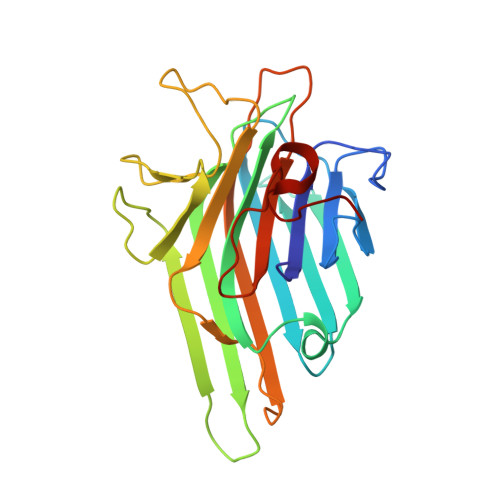
    Diocleinae lectins are highly homologous in their primary structure which features metal binding sites and a carbohydrate recognition domain (CRD). Differences in the biological activity of legume lectins have been widely investigated using hemagglutination inhibition assays, isothermal titration microcalorimetry and co-crystallization with mono- and oligosaccharides. Here we report a new lectin crystal structure (ConBr) extracted from seeds of Canavalia brasiliensis, predict dimannoside binding by docking, identify the α-aminobutyric acid (Abu) binding pocket and compare the CRD of ConBr to that of homologous lectins. Based on the hypothesis that the carbohydrate affinity of lectins depends on CRD configuration, the relationship between tridimensional structure and endothelial NO synthase activation was used to clarify differences in biological activity. Our study established a correlation between the position of CRD amino acid side chains and the stimulation of NO release from endothelium.


  • Organizational Affiliation

    Departamento de Bioquímica e Biologia Molecular, Universidade Federal do Ceará, Fortaleza, CE, Brazil.


Macromolecules
Find similar proteins by:  (by identity cutoff)  |  3D Structure
Entity ID: 1
MoleculeChains Sequence LengthOrganismDetailsImage
Concanavalin-Br237Canavalia brasiliensisMutation(s): 0 
UniProt
Find proteins for P55915 (Canavalia brasiliensis)
Explore P55915 
Go to UniProtKB:  P55915
Entity Groups  
Sequence Clusters30% Identity50% Identity70% Identity90% Identity95% Identity100% Identity
UniProt GroupP55915
Sequence Annotations
Expand
  • Reference Sequence
Small Molecules
Ligands 5 Unique
IDChains Name / Formula / InChI Key2D Diagram3D Interactions
DBB
Query on DBB

Download Ideal Coordinates CCD File 
B [auth A]D-ALPHA-AMINOBUTYRIC ACID
C4 H9 N O2
QWCKQJZIFLGMSD-GSVOUGTGSA-N
GOL
Query on GOL

Download Ideal Coordinates CCD File 
C [auth A]GLYCEROL
C3 H8 O3
PEDCQBHIVMGVHV-UHFFFAOYSA-N
MN
Query on MN

Download Ideal Coordinates CCD File 
E [auth A]MANGANESE (II) ION
Mn
WAEMQWOKJMHJLA-UHFFFAOYSA-N
CA
Query on CA

Download Ideal Coordinates CCD File 
D [auth A]CALCIUM ION
Ca
BHPQYMZQTOCNFJ-UHFFFAOYSA-N
CL
Query on CL

Download Ideal Coordinates CCD File 
F [auth A],
G [auth A]
CHLORIDE ION
Cl
VEXZGXHMUGYJMC-UHFFFAOYSA-M
Experimental Data & Validation

Experimental Data

  • Method: X-RAY DIFFRACTION
  • Resolution: 2.10 Å
  • R-Value Free: 0.253 
  • R-Value Work: 0.204 
  • R-Value Observed: 0.207 
  • Space Group: I 2 2 2
Unit Cell:
Length ( Å )Angle ( ˚ )
a = 68.323α = 90
b = 73.02β = 90
c = 99.542γ = 90
Software Package:
Software NamePurpose
HKL-2000data collection
MOLREPphasing
REFMACrefinement
MOSFLMdata reduction
SCALAdata scaling

Structure Validation

View Full Validation Report


Entry History 

Revision History  (Full details and data files)

  • Version 1.0: 2010-12-08
    Type: Initial release
  • Version 1.1: 2011-07-13
    Changes: Version format compliance
  • Version 1.2: 2023-09-06
    Changes: Data collection, Database references, Derived calculations, Refinement description
  • Version 1.3: 2023-11-22
    Changes: Data collection