3J3Z

Structure of MA28-7 neutralizing antibody Fab fragment from electron cryo-microscopy of enterovirus 71 complexed with a Fab fragment


Experimental Data Snapshot

  • Method: ELECTRON MICROSCOPY
  • Resolution: 23.4 Å
  • Aggregation State: PARTICLE 
  • Reconstruction Method: SINGLE PARTICLE 

wwPDB Validation   3D Report Full Report


This is version 1.3 of the entry. See complete history


Literature

A strain-specific epitope of enterovirus 71 identified by cryo-electron microscopy of the complex with fab from neutralizing antibody.

Lee, H.Cifuente, J.O.Ashley, R.E.Conway, J.F.Makhov, A.M.Tano, Y.Shimizu, H.Nishimura, Y.Hafenstein, S.

(2013) J Virol 87: 11363-11370

  • DOI: https://doi.org/10.1128/JVI.01926-13
  • Primary Citation of Related Structures:  
    3J3Z

  • PubMed Abstract: 

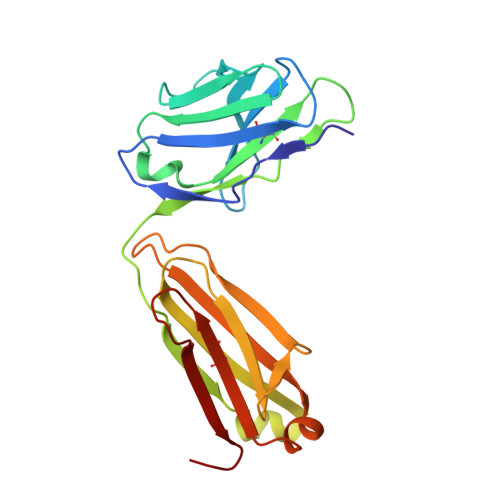
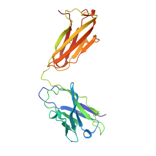
    Enterovirus 71 (EV71) is a picornavirus that causes outbreaks of hand, foot, and mouth disease (HFMD), primarily in the Asia-Pacific area. Unlike coxsackievirus A16, which also causes HFMD, EV71 induces severe neuropathology leading to high fatalities, especially among children under the age of 6 years. Currently, no established vaccines or treatments are available against EV71 infection. The monoclonal antibody MA28-7 neutralizes only specific strains of EV71 that have a conserved glycine at amino acid VP1-145, a surface-exposed residue that maps to the 5-fold vertex and that has been implicated in receptor binding. The cryo-electron microscopy structure of a complex between EV71 and the Fab fragment of MA28-7 shows that only one Fab fragment occupies each 5-fold vertex. A positively charged patch, which has also been implicated in receptor binding, lies within the Fab footprint. We identify the strain-specific epitope of EV71 and discuss the possible neutralization mechanisms of the antibody.


  • Organizational Affiliation

    Department of Medicine, The Pennsylvania State University College of Medicine, Hershey, Pennsylvania, USA.


Macromolecules
Find similar proteins by:  (by identity cutoff)  |  3D Structure
Entity ID: 1
MoleculeChains Sequence LengthOrganismDetailsImage
MA28-7 neutralizing antibody light chainA [auth L]214Mus musculusMutation(s): 0 
Entity Groups  
Sequence Clusters30% Identity50% Identity70% Identity90% Identity95% Identity100% Identity
Sequence Annotations
Expand
  • Reference Sequence
Find similar proteins by:  (by identity cutoff)  |  3D Structure
Entity ID: 2
MoleculeChains Sequence LengthOrganismDetailsImage
MA28-7 neutralizing antibody heavy chainB [auth H]220Mus musculusMutation(s): 0 
Entity Groups  
Sequence Clusters30% Identity50% Identity70% Identity90% Identity95% Identity100% Identity
Sequence Annotations
Expand
  • Reference Sequence
Experimental Data & Validation

Experimental Data

  • Method: ELECTRON MICROSCOPY
  • Resolution: 23.4 Å
  • Aggregation State: PARTICLE 
  • Reconstruction Method: SINGLE PARTICLE 
EM Software:
TaskSoftware PackageVersion
RECONSTRUCTIONEMAN2

Structure Validation

View Full Validation Report


Entry History 

Deposition Data

Revision History  (Full details and data files)

  • Version 1.0: 2013-08-28
    Type: Initial release
  • Version 1.1: 2013-11-06
    Changes: Database references
  • Version 1.2: 2014-04-09
    Changes: Source and taxonomy
  • Version 1.3: 2018-07-18
    Changes: Data collection