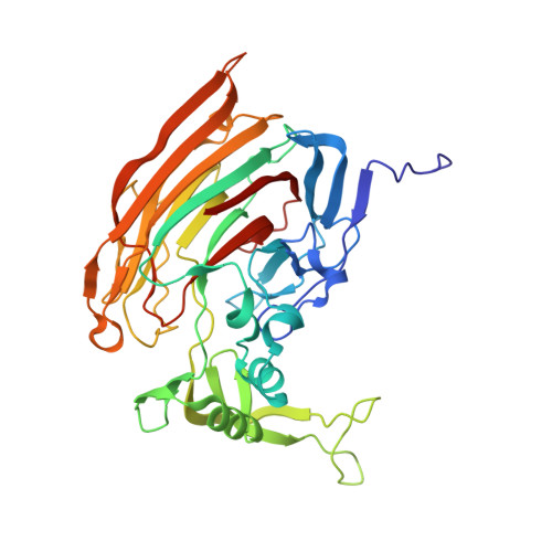
3IKW

Structure of Heparinase I from Bacteroides thetaiotaomicron


Experimental Data Snapshot

  • Method: X-RAY DIFFRACTION
  • Resolution: 1.30 Å
  • R-Value Free: 0.210 
  • R-Value Work: 0.187 
  • R-Value Observed: 0.188 

wwPDB Validation   3D Report Full Report


This is version 1.3 of the entry. See complete history


Literature

Structural snapshots of heparin depolymerization by heparin lyase I.

Han, Y.H.Garron, M.L.Kim, H.Y.Kim, W.S.Zhang, Z.Ryu, K.S.Shaya, D.Xiao, Z.Cheong, C.Kim, Y.S.Linhardt, R.J.Jeon, Y.H.Cygler, M.

(2009) J Biol Chem 284: 34019-34027

  • DOI: https://doi.org/10.1074/jbc.M109.025338
  • Primary Citation of Related Structures:  
    3IKW, 3ILR, 3IMN, 3IN9, 3INA

  • PubMed Abstract: 

    Heparin lyase I (heparinase I) specifically depolymerizes heparin, cleaving the glycosidic linkage next to iduronic acid. Here, we show the crystal structures of heparinase I from Bacteroides thetaiotaomicron at various stages of the reaction with heparin oligosaccharides before and just after cleavage and product disaccharide. The heparinase I structure is comprised of a beta-jellyroll domain harboring a long and deep substrate binding groove and an unusual thumb-resembling extension. This thumb, decorated with many basic residues, is of particular importance in activity especially on short heparin oligosaccharides. Unexpected structural similarity of the active site to that of heparinase II with an (alpha/alpha)(6) fold is observed. Mutational studies and kinetic analysis of this enzyme provide insights into the catalytic mechanism, the substrate recognition, and processivity.


  • Organizational Affiliation

    Magnetic Resonance Team, Korea Basic Science Institute, Ochang, Chungbuk 363-883, Korea.


Macromolecules
Find similar proteins by:  (by identity cutoff)  |  3D Structure
Entity ID: 1
MoleculeChains Sequence LengthOrganismDetailsImage
Heparin lyase I374Bacteroides thetaiotaomicronMutation(s): 0 
Gene Names: BT_4675
EC: 4.2.2.7
UniProt
Find proteins for Q89YQ6 (Bacteroides thetaiotaomicron (strain ATCC 29148 / DSM 2079 / JCM 5827 / CCUG 10774 / NCTC 10582 / VPI-5482 / E50))
Explore Q89YQ6 
Go to UniProtKB:  Q89YQ6
Entity Groups  
Sequence Clusters30% Identity50% Identity70% Identity90% Identity95% Identity100% Identity
UniProt GroupQ89YQ6
Sequence Annotations
Expand
  • Reference Sequence
Experimental Data & Validation

Experimental Data

  • Method: X-RAY DIFFRACTION
  • Resolution: 1.30 Å
  • R-Value Free: 0.210 
  • R-Value Work: 0.187 
  • R-Value Observed: 0.188 
  • Space Group: P 21 21 2
Unit Cell:
Length ( Å )Angle ( ˚ )
a = 76.263α = 90
b = 110.494β = 90
c = 44.158γ = 90
Software Package:
Software NamePurpose
DENZOdata reduction
SCALEPACKdata scaling
SHELXphasing
REFMACrefinement
PDB_EXTRACTdata extraction
HKL-2000data collection
SHELXDphasing

Structure Validation

View Full Validation Report


Entry History 

Deposition Data

Revision History  (Full details and data files)

  • Version 1.0: 2009-09-29
    Type: Initial release
  • Version 1.1: 2011-07-13
    Changes: Version format compliance
  • Version 1.2: 2017-11-01
    Changes: Refinement description
  • Version 1.3: 2024-02-21
    Changes: Data collection, Database references, Derived calculations