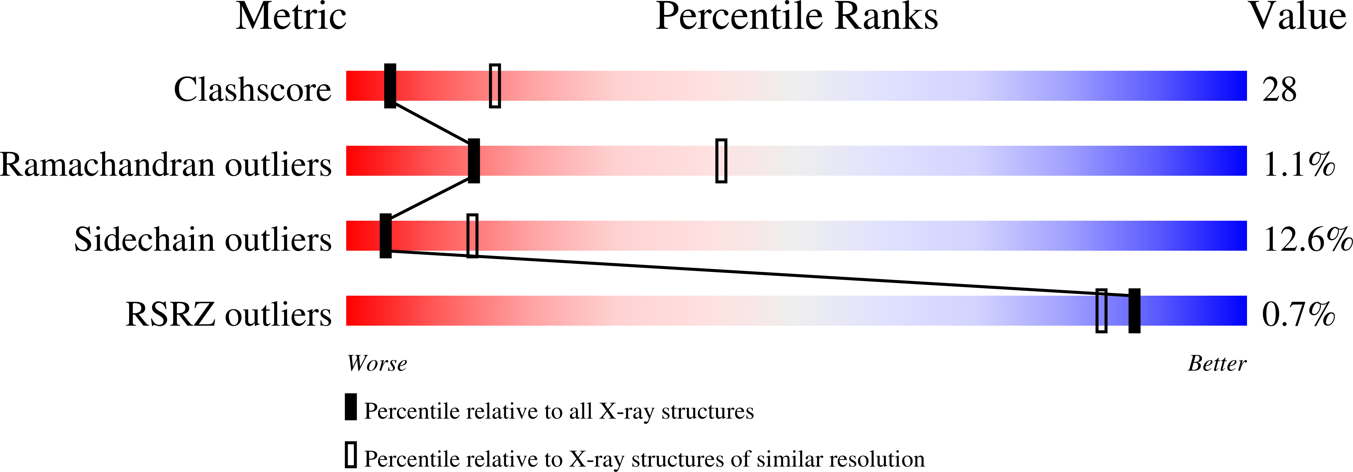
Conformational plasticity of the coiled-coil domain of BmrR is required for bmr operator binding: the structure of unliganded BmrR.
Kumaraswami, M., Newberry, K.J., Brennan, R.G.(2010) J Mol Biol 398: 264-275
- PubMed: 20230832
- DOI: https://doi.org/10.1016/j.jmb.2010.03.011
- Primary Citation of Related Structures:
3IAO - PubMed Abstract:
The multidrug-binding transcription regulator BmrR from Bacillus subtilis is a MerR family member that binds to a wide array of cationic lipophilic toxins to activate the transcription of the multidrug efflux pump gene bmr. Transcription activation from the sigma(A)-dependent bmr operator requires BmrR to remodel the nonoptimal 19-bp spacer between the -10 promoter element and the -35 promoter element in order to facilitate productive RNA polymerase binding. Despite the availability of several structures of BmrR bound to DNA and drugs, the lack of a BmrR structure in its unliganded or apo (DNA free and drug free) state hinders our full understanding of the structural transitions required for DNA binding and transcription activation. Here, we report the crystal structure of the constitutively active, unliganded BmrR mutant BmrR(E253Q/R275E). Superposition of the ligand-free (apo BmrR(E253Q/R275E)) and DNA-bound BmrR structures reveals that apo BmrR must undergo significant rearrangement in order to assume the DNA-bound conformation, including an outward rotation of minor groove binding wings, an inward movement of helix-turn-helix motifs, and a downward relocation of pliable coiled-coil helices. Computational analysis of the DNA-free and DNA-bound structures reveals a flexible joint that is located at the center of the coiled-coil helices. This region, which is composed of residues 94 through 98, overlaps the helical bulge that is observed only in the apo BmrR structure. This conformational hinge is likely common to other MerR family members with large effector-binding domains, but appears to be missing from the smaller metal-binding MerR family members. Interestingly, the center-to-center distance of the recognition helices of apo BmrR is 34 A and suggests that the conformational change from the apo BmrR structure to the bmr operator-bound BmrR structure is initiated by the binding of this transcription activator to a more B-DNA-like conformation.
Organizational Affiliation:
Department of Biochemistry and Molecular Biology, Center for Biomolecular Structure and Function, The University of Texas M. D. Anderson Cancer Center, Houston, TX 77030, USA.


