3I39

NI,FE-CODH-320 MV+CN state


Experimental Data Snapshot

  • Method: X-RAY DIFFRACTION
  • Resolution: 1.36 Å
  • R-Value Free: 0.190 
  • R-Value Work: 0.154 
  • R-Value Observed: 0.156 

wwPDB Validation   3D Report Full Report


This is version 1.4 of the entry. See complete history


Literature

Structural basis of cyanide inhibition of Ni, Fe-containing carbon monoxide dehydrogenase

Jeoung, J.H.Dobbek, H.

(2009) J Am Chem Soc 131: 9922-9923

  • DOI: https://doi.org/10.1021/ja9046476
  • Primary Citation of Related Structures:  
    3I39

  • PubMed Abstract: 

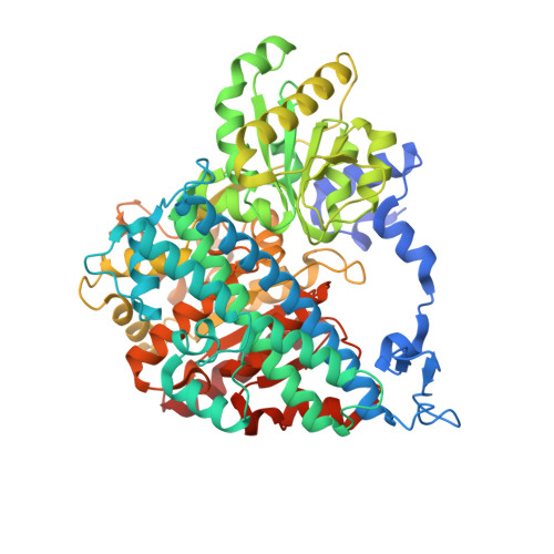
    Carbon monoxide dehydrogenases (CODHs) catalyze the reversible oxidation of carbon monoxide with water to carbon dioxide, two protons, and two electrons. The CODHs of anaerobic microorganisms harbor a complex Ni/Fe/S-containing metal center called a C-cluster in their active site, which activates the substrates water and carbon monoxide, stabilizes an intermediary metal-carboxylate, and transiently stores the two electrons generated in the reaction. Several small molecules have been reported to inhibit carbon monoxide oxidation by CODHs, among which the cyanide anion acts as a slow binding inhibitor. Cyanide is isoelectronic to the substrate carbon monoxide, and its binding to the C-cluster has been reported to involve nickel, nickel and iron, or only iron. We report the crystal structure of CODH-II from Carboxydothermus hydrogenoformans in complex with cyanide at 1.36 A resolution. The structure reveals that cyanide binds to the C-cluster at an open coordination site completing the square-planar coordination geometry of the nickel ion. While active CODH has a water/hydroxo-ligand bound to an iron ion near nickel, in the cyanide complex the water/hydroxo-ligand is lost and iron occupies a position more close to the nickel ion. Based on the structure, we suggest that the competitive inhibitory character of cyanide originates from it obstruction of carbon monoxide binding to the nickel ion while the slow binding inhibition is due to a conformational change of the protein during which the water/hydroxo-ligand bound to iron is lost.


  • Organizational Affiliation

    Labor für Bioanorganische Chemie, Universität Bayreuth, 95447 Bayreuth, Germany.


Macromolecules
Find similar proteins by:  (by identity cutoff)  |  3D Structure
Entity ID: 1
MoleculeChains Sequence LengthOrganismDetailsImage
Carbon monoxide dehydrogenase 2A [auth X]656Carboxydothermus hydrogenoformans Z-2901Mutation(s): 1 
Gene Names: CHY_0085cooS2cooSII
EC: 1.2.99.2
UniProt
Find proteins for Q9F8A8 (Carboxydothermus hydrogenoformans (strain ATCC BAA-161 / DSM 6008 / Z-2901))
Explore Q9F8A8 
Go to UniProtKB:  Q9F8A8
Entity Groups  
Sequence Clusters30% Identity50% Identity70% Identity90% Identity95% Identity100% Identity
UniProt GroupQ9F8A8
Sequence Annotations
Expand
  • Reference Sequence
Small Molecules
Ligands 5 Unique
IDChains Name / Formula / InChI Key2D Diagram3D Interactions
WCC
Query on WCC

Download Ideal Coordinates CCD File 
D [auth X]FE(3)-NI(1)-S(4) CLUSTER
Fe3 Ni S4
SEEZYPKDPRYISB-UHFFFAOYSA-N
SF4
Query on SF4

Download Ideal Coordinates CCD File 
B [auth X]IRON/SULFUR CLUSTER
Fe4 S4
LJBDFODJNLIPKO-UHFFFAOYSA-N
FES
Query on FES

Download Ideal Coordinates CCD File 
C [auth X]FE2/S2 (INORGANIC) CLUSTER
Fe2 S2
NIXDOXVAJZFRNF-UHFFFAOYSA-N
FE2
Query on FE2

Download Ideal Coordinates CCD File 
E [auth X],
F [auth X]
FE (II) ION
Fe
CWYNVVGOOAEACU-UHFFFAOYSA-N
CYN
Query on CYN

Download Ideal Coordinates CCD File 
G [auth X]CYANIDE ION
C N
XFXPMWWXUTWYJX-UHFFFAOYSA-N
Experimental Data & Validation

Experimental Data

  • Method: X-RAY DIFFRACTION
  • Resolution: 1.36 Å
  • R-Value Free: 0.190 
  • R-Value Work: 0.154 
  • R-Value Observed: 0.156 
  • Space Group: C 1 2 1
Unit Cell:
Length ( Å )Angle ( ˚ )
a = 111.279α = 90
b = 75.834β = 111.33
c = 71.099γ = 90
Software Package:
Software NamePurpose
REFMACrefinement
CNSrefinement
MAR345dtbdata collection
XDSdata reduction
XSCALEdata scaling
CNSphasing

Structure Validation

View Full Validation Report


Entry History 

Deposition Data

Revision History  (Full details and data files)

  • Version 1.0: 2009-08-11
    Type: Initial release
  • Version 1.1: 2011-07-13
    Changes: Source and taxonomy, Version format compliance
  • Version 1.2: 2019-07-24
    Changes: Data collection, Refinement description
  • Version 1.3: 2021-10-13
    Changes: Database references, Derived calculations
  • Version 1.4: 2024-02-21
    Changes: Data collection