3HUB

Human p38 MAP Kinase in Complex with Scios-469


Experimental Data Snapshot

  • Method: X-RAY DIFFRACTION
  • Resolution: 2.25 Å
  • R-Value Free: 0.292 
  • R-Value Work: 0.213 
  • R-Value Observed: 0.217 

wwPDB Validation   3D Report Full Report


This is version 1.4 of the entry. See complete history


Literature

Fluorophore labeling of the glycine-rich loop as a method of identifying inhibitors that bind to active and inactive kinase conformations.

Simard, J.R.Getlik, M.Grutter, C.Schneider, R.Wulfert, S.Rauh, D.

(2010) J Am Chem Soc 132: 4152-4160

  • DOI: https://doi.org/10.1021/ja908083e
  • Primary Citation of Related Structures:  
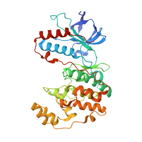
    3HUB, 3HUC, 3L8S

  • PubMed Abstract: 

    Targeting protein kinases with small organic molecules is a promising strategy to regulate unwanted kinase activity in both chemical biology and medicinal chemistry research. Traditionally, kinase inhibitors are identified in activity-based screening assays using enzymatically active kinase preparations to measure the perturbation of substrate phosphorylation, often resulting in the enrichment of classical ATP competitive (Type I) inhibitors. However, addressing enzymatically incompetent kinase conformations offers new opportunities for targeted therapies and is moving to the forefront of kinase inhibitor research. Here we report the development of a new FLiK (Fluorescent Labels in Kinases) binding assay to detect small molecules that induce changes in the conformation of the glycine-rich loop. Due to cross-talk between the glycine-rich loop and the activation loop in kinases, this alternative labeling approach can also detect ligands that stabilize inactive kinase conformations, including slow-binding Type II and Type III kinase inhibitors. Protein X-ray crystallography validated the assay results and identified a novel DFG-out binding mode for a quinazoline-based inhibitor in p38alpha kinase. We also detected the high-affinity binding of a clinically relevant and specific VEGFR2 inhibitor, and we provide structural details of its binding mode in p38alpha, in which it stabilizes the DFG-out conformation. Last, we demonstrate the power of this new FLiK labeling strategy to detect the binding of Type I ligands that induce conformational changes in the glycine-rich loop as a means of gaining affinity for the target kinase. This approach may be a useful alternative to develop direct binding assays for kinases that do not adopt the DFG-out conformation while also avoiding the use of expensive kits, detection reagents, or radioactivity frequently employed with activity-based assays.


  • Organizational Affiliation

    Chemical Genomics Centre of the Max Planck Society, Otto-Hahn-Strasse 15, D-44227 Dortmund, Germany.


Macromolecules
Find similar proteins by:  (by identity cutoff)  |  3D Structure
Entity ID: 1
MoleculeChains Sequence LengthOrganismDetailsImage
Mitogen-activated protein kinase 14360Homo sapiensMutation(s): 4 
Gene Names: MAPK14CSBPCSBP1CSBP2CSPB1MXI2
EC: 2.7.11.24
UniProt & NIH Common Fund Data Resources
Find proteins for Q16539 (Homo sapiens)
Explore Q16539 
Go to UniProtKB:  Q16539
PHAROS:  Q16539
GTEx:  ENSG00000112062 
Entity Groups  
Sequence Clusters30% Identity50% Identity70% Identity90% Identity95% Identity100% Identity
UniProt GroupQ16539
Sequence Annotations
Expand
  • Reference Sequence
Small Molecules
Ligands 2 Unique
IDChains Name / Formula / InChI Key2D Diagram3D Interactions
469
Query on 469

Download Ideal Coordinates CCD File 
C [auth A]2-(6-chloro-5-{[(2R,5S)-4-(4-fluorobenzyl)-2,5-dimethylpiperazin-1-yl]carbonyl}-1-methyl-1H-indol-3-yl)-N,N-dimethyl-2-oxoacetamide
C27 H30 Cl F N4 O3
ZMELOYOKMZBMRB-DLBZAZTESA-N
BOG
Query on BOG

Download Ideal Coordinates CCD File 
B [auth A]octyl beta-D-glucopyranoside
C14 H28 O6
HEGSGKPQLMEBJL-RKQHYHRCSA-N
Binding Affinity Annotations 
IDSourceBinding Affinity
469 BindingDB:  3HUB Kd: 0.48 (nM) from 1 assay(s)
IC50: min: 9, max: 96 (nM) from 4 assay(s)
Binding MOAD:  3HUB Kd: 8.2 (nM) from 1 assay(s)
PDBBind:  3HUB Kd: 8.2 (nM) from 1 assay(s)
Experimental Data & Validation

Experimental Data

  • Method: X-RAY DIFFRACTION
  • Resolution: 2.25 Å
  • R-Value Free: 0.292 
  • R-Value Work: 0.213 
  • R-Value Observed: 0.217 
  • Space Group: P 21 21 21
Unit Cell:
Length ( Å )Angle ( ˚ )
a = 68.01α = 90
b = 69.61β = 90
c = 74.44γ = 90
Software Package:
Software NamePurpose
XSCALEdata scaling
PHASERphasing
REFMACrefinement
PDB_EXTRACTdata extraction
XDSdata scaling
XDSdata reduction

Structure Validation

View Full Validation Report


Entry History 

Deposition Data

Revision History  (Full details and data files)

  • Version 1.0: 2010-03-09
    Type: Initial release
  • Version 1.1: 2011-07-13
    Changes: Version format compliance
  • Version 1.2: 2020-07-29
    Type: Remediation
    Reason: Carbohydrate remediation
    Changes: Data collection, Database references, Derived calculations, Structure summary
  • Version 1.3: 2021-10-13
    Changes: Database references, Structure summary
  • Version 1.4: 2024-02-21
    Changes: Data collection