3H1O

The Structure of Fluorescent Protein FP480


Experimental Data Snapshot

  • Method: X-RAY DIFFRACTION
  • Resolution: 2.00 Å
  • R-Value Free: 0.220 
  • R-Value Work: 0.175 

wwPDB Validation   3D Report Full Report


This is version 1.2 of the entry. See complete history


Literature

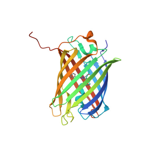
Rotational order-disorder structure of fluorescent protein FP480

Pletnev, S.Morozova, K.S.Verkhusha, V.V.Dauter, Z.

(2009) Acta Crystallogr D Biol Crystallogr 65: 906-912

  • DOI: https://doi.org/10.1107/S0907444909020927
  • Primary Citation of Related Structures:  
    3H1O, 3H1R

  • PubMed Abstract: 

    In the last decade, advances in instrumentation and software development have made crystallography a powerful tool in structural biology. Using this method, structural information can now be acquired from pathological crystals that would have been abandoned in earlier times. In this paper, the order-disorder (OD) structure of fluorescent protein FP480 is discussed. The structure is composed of tetramers with 222 symmetry incorporated into the lattice in two different ways, namely rotated 90 degrees with respect to each other around the crystal c axis, with tetramer axes coincident with crystallographic twofold axes. The random distribution of alternatively oriented tetramers in the crystal creates a rotational OD structure with statistically averaged I422 symmetry, although the presence of very weak and diffuse additional reflections suggests that the randomness is only approximate.


  • Organizational Affiliation

    SAIC-Frederick Inc., Basic Research Program, Argonne National Laboratory, Argonne, IL 60439, USA. svp@ncifcrf.gov


Macromolecules
Find similar proteins by:  (by identity cutoff)  |  3D Structure
Entity ID: 1
MoleculeChains Sequence LengthOrganismDetailsImage
Fluorescent protein FP480
A, B
231Entacmaea quadricolorMutation(s): 0 
UniProt
Find proteins for D0VX33 (Entacmaea quadricolor)
Explore D0VX33 
Go to UniProtKB:  D0VX33
Entity Groups  
Sequence Clusters30% Identity50% Identity70% Identity90% Identity95% Identity100% Identity
UniProt GroupD0VX33
Sequence Annotations
Expand
  • Reference Sequence
Experimental Data & Validation

Experimental Data

  • Method: X-RAY DIFFRACTION
  • Resolution: 2.00 Å
  • R-Value Free: 0.220 
  • R-Value Work: 0.175 
  • Space Group: I 4
Unit Cell:
Length ( Å )Angle ( ˚ )
a = 98.208α = 90
b = 98.208β = 90
c = 108.058γ = 90
Software Package:
Software NamePurpose
DENZOdata reduction
SCALEPACKdata scaling
PHENIXrefinement
PDB_EXTRACTdata extraction
SERGUIdata collection
MOLREPphasing

Structure Validation

View Full Validation Report


Entry History 

Deposition Data

Revision History  (Full details and data files)

  • Version 1.0: 2009-09-08
    Type: Initial release
  • Version 1.1: 2011-07-13
    Changes: Non-polymer description, Version format compliance
  • Version 1.2: 2017-11-01
    Changes: Refinement description