3GW1

The structure of the Caulobacter crescentus CLPs protease adaptor protein in complex with FGG tripeptide


Experimental Data Snapshot

  • Method: X-RAY DIFFRACTION
  • Resolution: 2.36 Å
  • R-Value Free: 0.297 
  • R-Value Work: 0.270 
  • R-Value Observed: 0.272 

wwPDB Validation   3D Report Full Report


This is version 1.3 of the entry. See complete history


Literature

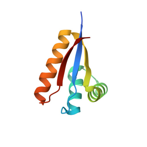
Molecular basis of substrate selection by the N-end rule adaptor protein ClpS.

Roman-Hernandez, G.Grant, R.A.Sauer, R.T.Baker, T.A.

(2009) Proc Natl Acad Sci U S A 106: 8888-8893

  • DOI: https://doi.org/10.1073/pnas.0903614106
  • Primary Citation of Related Structures:  
    3G19, 3G1B, 3GQ0, 3GQ1, 3GW1

  • PubMed Abstract: 

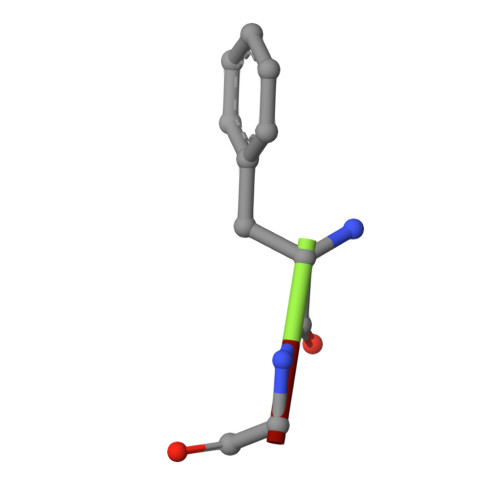
    The N-end rule is a conserved degradation pathway that relates the stability of a protein to its N-terminal amino acid. Here, we present crystal structures of ClpS, the bacterial N-end rule adaptor, alone and engaged with peptides containing N-terminal phenylalanine, leucine, and tryptophan. These structures, together with a previous structure of ClpS bound to an N-terminal tyrosine, illustrate the molecular basis of recognition of the complete set of primary N-end rule amino acids. In each case, the alpha-amino group and side chain of the N-terminal residue are the major determinants of recognition. The binding pocket for the N-end residue is preformed in the free adaptor, and only small adjustments are needed to accommodate N-end rule residues having substantially different sizes and shapes. M53A ClpS is known to mediate degradation of an expanded repertoire of substrates, including those with N-terminal valine or isoleucine. A structure of Met53A ClpS engaged with an N-end rule tryptophan reveals an essentially wild-type mechanism of recognition, indicating that the Met(53) side chain directly enforces specificity by clashing with and excluding beta-branched side chains. Finally, experimental and structural data suggest mechanisms that make proteins with N-terminal methionine bind very poorly to ClpS, explaining why these high-abundance proteins are not degraded via the N-end rule pathway in the cell.


  • Organizational Affiliation

    Department of Biology, Massachusetts Institute of Technology, Cambridge, MA 02139, USA.


Macromolecules
Find similar proteins by:  (by identity cutoff)  |  3D Structure
Entity ID: 1
MoleculeChains Sequence LengthOrganismDetailsImage
ATP-dependent Clp protease adapter protein ClpS
A, B
85Caulobacter vibrioidesMutation(s): 0 
Gene Names: clpS
UniProt
Find proteins for A0A290MK63 (Caulobacter vibrioides)
Explore A0A290MK63 
Go to UniProtKB:  A0A290MK63
Entity Groups  
Sequence Clusters30% Identity50% Identity70% Identity90% Identity95% Identity100% Identity
UniProt GroupA0A290MK63
Sequence Annotations
Expand
  • Reference Sequence

Find similar proteins by:  Sequence   |   3D Structure  

Entity ID: 2
MoleculeChains Sequence LengthOrganismDetailsImage
FGG peptide
C, D
3N/AMutation(s): 0 
Sequence Annotations
Expand
  • Reference Sequence
Small Molecules
Ligands 1 Unique
IDChains Name / Formula / InChI Key2D Diagram3D Interactions
MG
Query on MG

Download Ideal Coordinates CCD File 
E [auth A]MAGNESIUM ION
Mg
JLVVSXFLKOJNIY-UHFFFAOYSA-N
Experimental Data & Validation

Experimental Data

  • Method: X-RAY DIFFRACTION
  • Resolution: 2.36 Å
  • R-Value Free: 0.297 
  • R-Value Work: 0.270 
  • R-Value Observed: 0.272 
  • Space Group: P 1 21 1
Unit Cell:
Length ( Å )Angle ( ˚ )
a = 27.599α = 90
b = 38.32β = 90.1
c = 62.857γ = 90
Software Package:
Software NamePurpose
DENZOdata reduction
SCALEPACKdata scaling
PHENIXrefinement
PDB_EXTRACTdata extraction
HKL-2000data collection

Structure Validation

View Full Validation Report


Entry History 

Deposition Data

Revision History  (Full details and data files)

  • Version 1.0: 2009-05-05
    Type: Initial release
  • Version 1.1: 2011-07-13
    Changes: Version format compliance
  • Version 1.2: 2017-11-01
    Changes: Refinement description
  • Version 1.3: 2023-09-06
    Changes: Data collection, Database references, Refinement description, Source and taxonomy, Structure summary