3GTN

Crystal Structure of XynC from Bacillus subtilis 168


Experimental Data Snapshot

  • Method: X-RAY DIFFRACTION
  • Resolution: 2.68 Å
  • R-Value Free: 0.287 
  • R-Value Work: 0.236 
  • R-Value Observed: 0.239 

wwPDB Validation   3D Report Full Report


This is version 1.3 of the entry. See complete history


Literature

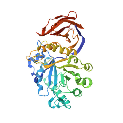
Crystallization and crystallographic analysis of Bacillus subtilis xylanase C.

St John, F.J.Godwin, D.K.Preston, J.F.Pozharski, E.Hurlbert, J.C.

(2009) Acta Crystallogr Sect F Struct Biol Cryst Commun 65: 499-503

  • DOI: https://doi.org/10.1107/S1744309109013098
  • Primary Citation of Related Structures:  
    3GTN

  • PubMed Abstract: 

    The recent biochemical characterization of the xylanases of glycosyl hydrolase family 5 (GH 5) has identified a distinctive endo mode of action, hydrolyzing the beta-1,4 xylan chain at a specific site directed by the position of an alpha-1,2-linked glucuronate moiety. Xylanase C (XynC), the GH 5 xylanase from Bacillus subtilis 168, has been cloned, overexpressed and crystallized. Initial data collection was performed and a preliminary model has been built into a low-quality 2.7 A resolution density map. The crystals belonged to the primitive monoclinic space group P2(1). Further screening identified an additive that resulted in large reproducible crystals. This larger more robust crystal form belonged to space group P2(1)2(1)2 and a resulting data set has been processed to 1.64 A resolution. This will be the second structure to be solved from this unique xylanase family and the first from a Gram-positive bacterium. This work may help to identify the structural determinants that allow the exceptional specificity of this enzyme and the role it plays in the biological depolymerization and processing of glucuronoxylan.


  • Organizational Affiliation

    University of Maryland School of Pharmacy, Department of Pharmaceutical Sciences, Baltimore, 21201, USA. fjstjohn@gmail.com


Macromolecules
Find similar proteins by:  (by identity cutoff)  |  3D Structure
Entity ID: 1
MoleculeChains Sequence LengthOrganismDetailsImage
Glucuronoxylanase xynC
A, B
401Bacillus subtilisMutation(s): 0 
Gene Names: BSU18150xynCynfF
EC: 3.2.1.136
UniProt
Find proteins for Q45070 (Bacillus subtilis (strain 168))
Explore Q45070 
Go to UniProtKB:  Q45070
Entity Groups  
Sequence Clusters30% Identity50% Identity70% Identity90% Identity95% Identity100% Identity
UniProt GroupQ45070
Sequence Annotations
Expand
  • Reference Sequence
Experimental Data & Validation

Experimental Data

  • Method: X-RAY DIFFRACTION
  • Resolution: 2.68 Å
  • R-Value Free: 0.287 
  • R-Value Work: 0.236 
  • R-Value Observed: 0.239 
  • Space Group: P 1 21 1
Unit Cell:
Length ( Å )Angle ( ˚ )
a = 61.338α = 90
b = 82.108β = 104.7
c = 96.648γ = 90
Software Package:
Software NamePurpose
DENZOdata reduction
SCALEPACKdata scaling
PHASERphasing
REFMACrefinement
PDB_EXTRACTdata extraction
Blu-Icedata collection
HKL-2000data reduction
HKL-2000data scaling

Structure Validation

View Full Validation Report


Entry History 

Deposition Data

Revision History  (Full details and data files)

  • Version 1.0: 2009-04-28
    Type: Initial release
  • Version 1.1: 2011-07-13
    Changes: Version format compliance
  • Version 1.2: 2017-11-01
    Changes: Refinement description
  • Version 1.3: 2024-02-21
    Changes: Data collection, Database references, Refinement description