3GMB

Crystal Structure of 2-Methyl-3-hydroxypyridine-5-carboxylic acid Oxygenase


Experimental Data Snapshot

  • Method: X-RAY DIFFRACTION
  • Resolution: 2.10 Å
  • R-Value Free: 0.224 
  • R-Value Work: 0.193 
  • R-Value Observed: 0.193 

wwPDB Validation   3D Report Full Report


Ligand Structure Quality Assessment 


This is version 1.3 of the entry. See complete history


Literature

Structure of the PLP degradative enzyme 2-methyl-3-hydroxypyridine-5-carboxylic acid oxygenase from Mesorhizobium loti MAFF303099 and its mechanistic implications.

McCulloch, K.M.Mukherjee, T.Begley, T.P.Ealick, S.E.

(2009) Biochemistry 48: 4139-4149

  • DOI: https://doi.org/10.1021/bi900149f
  • Primary Citation of Related Structures:  
    3GMB, 3GMC

  • PubMed Abstract: 

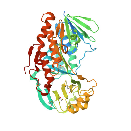
    A vitamin B(6) degradative pathway has recently been identified and characterized in Mesorhizobium loti MAFF303099. One of the enzymes on this pathway, 2-methyl-3-hydroxypyridine-5-carboxylic acid oxygenase (MHPCO), is a flavin-dependent enzyme and catalyzes the oxidative ring-opening of 2-methyl-3-hydroxypyridine-5-carboxylic acid to form E-2-(acetamino-methylene)succinate. The gene for this enzyme has been cloned, and the corresponding protein has been overexpressed in Escherichia coli and purified. The crystal structure of MHPCO has been solved to 2.1 A using SAD phasing with and without the substrate MHPC bound. These crystal structures provide insight into the reaction mechanism and suggest roles for active site residues in the catalysis of a novel oxidative ring-opening reaction.


  • Organizational Affiliation

    Department of Chemistry and Chemical Biology, Cornell University, Ithaca, New York 14853, USA.


Macromolecules
Find similar proteins by:  (by identity cutoff)  |  3D Structure
Entity ID: 1
MoleculeChains Sequence LengthOrganismDetailsImage
2-methyl-3-hydroxypyridine-5-carboxylic acid oxygenase
A, B
415Mesorhizobium lotiMutation(s): 0 
Gene Names: mlr6788
UniProt
Find proteins for Q988D3 (Mesorhizobium japonicum (strain LMG 29417 / CECT 9101 / MAFF 303099))
Explore Q988D3 
Go to UniProtKB:  Q988D3
Entity Groups  
Sequence Clusters30% Identity50% Identity70% Identity90% Identity95% Identity100% Identity
UniProt GroupQ988D3
Sequence Annotations
Expand
  • Reference Sequence
Small Molecules
Ligands 1 Unique
IDChains Name / Formula / InChI Key2D Diagram3D Interactions
FAD
Query on FAD

Download Ideal Coordinates CCD File 
C [auth A],
D [auth B]
FLAVIN-ADENINE DINUCLEOTIDE
C27 H33 N9 O15 P2
VWWQXMAJTJZDQX-UYBVJOGSSA-N
Modified Residues  1 Unique
IDChains TypeFormula2D DiagramParent
MSE
Query on MSE
A, B
L-PEPTIDE LINKINGC5 H11 N O2 SeMET
Experimental Data & Validation

Experimental Data

  • Method: X-RAY DIFFRACTION
  • Resolution: 2.10 Å
  • R-Value Free: 0.224 
  • R-Value Work: 0.193 
  • R-Value Observed: 0.193 
  • Space Group: C 1 2 1
Unit Cell:
Length ( Å )Angle ( ˚ )
a = 111.28α = 90
b = 130.2β = 122.6
c = 89.54γ = 90
Software Package:
Software NamePurpose
DENZOdata reduction
SCALEPACKdata scaling
CNSrefinement
PDB_EXTRACTdata extraction
HKL-2000data collection
HKL-2000data reduction
CNSphasing

Structure Validation

View Full Validation Report


Ligand Structure Quality Assessment 


Entry History 

Deposition Data

Revision History  (Full details and data files)

  • Version 1.0: 2009-04-14
    Type: Initial release
  • Version 1.1: 2011-07-13
    Changes: Version format compliance
  • Version 1.2: 2012-12-26
    Changes: Other, Structure summary
  • Version 1.3: 2017-11-01
    Changes: Refinement description