


Crystal structure of N-ethylmaleimidine reductase from Burkholderia pseudomallei
Edwards, T.E., Staker, B.L., Seattle Structural Genomics Center for Infectious Disease (SSGCID)To be published.
Experimental Data Snapshot
Entity ID: 1 | |||||
|---|---|---|---|---|---|
| Molecule | Chains | Sequence Length | Organism | Details | Image |
| N-ethylmaleimide reductase | 361 | Burkholderia pseudomallei | Mutation(s): 0 Gene Names: BPSS0877 | 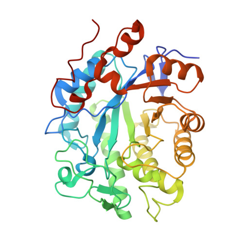 | |
UniProt | |||||
Find proteins for Q3JFM9 (Burkholderia pseudomallei (strain 1710b)) Explore Q3JFM9 Go to UniProtKB: Q3JFM9 | |||||
Entity Groups | |||||
| Sequence Clusters | 30% Identity50% Identity70% Identity90% Identity95% Identity100% Identity | ||||
| UniProt Group | Q3JFM9 | ||||
Sequence AnnotationsExpand | |||||
| |||||
| Ligands 1 Unique | |||||
|---|---|---|---|---|---|
| ID | Chains | Name / Formula / InChI Key | 2D Diagram | 3D Interactions | |
| FMN Query on FMN | C [auth A], D [auth B] | FLAVIN MONONUCLEOTIDE C17 H21 N4 O9 P FVTCRASFADXXNN-SCRDCRAPSA-N |  | ||
| Length ( Å ) | Angle ( ˚ ) |
|---|---|
| a = 49.35 | α = 90 |
| b = 77.89 | β = 90 |
| c = 168.94 | γ = 90 |
| Software Name | Purpose |
|---|---|
| XSCALE | data scaling |
| PHASER | phasing |
| REFMAC | refinement |
| PDB_EXTRACT | data extraction |
RCSB PDB (citation) is hosted by
RCSB PDB is a member of the

