


The crystal structure of gluconate kinase from Lactobacillus acidophilus
Zhang, Z., Swaminathan, S., Burley, S.K.To be published.
Experimental Data Snapshot
wwPDB Validation 3D Report Full Report

Entity ID: 1 | |||||
|---|---|---|---|---|---|
| Molecule | Chains | Sequence Length | Organism | Details | Image |
| Gluconate kinase | 504 | Lactobacillus acidophilus NCFM | Mutation(s): 0 Gene Names: LBA0354 EC: 2.7.1.12 | 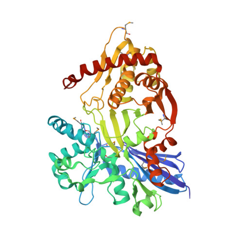 | |
UniProt | |||||
Find proteins for Q5FM28 (Lactobacillus acidophilus (strain ATCC 700396 / NCK56 / N2 / NCFM)) Explore Q5FM28 Go to UniProtKB: Q5FM28 | |||||
Entity Groups | |||||
| Sequence Clusters | 30% Identity50% Identity70% Identity90% Identity95% Identity100% Identity | ||||
| UniProt Group | Q5FM28 | ||||
Sequence AnnotationsExpand | |||||
| |||||
| Modified Residues 1 Unique | |||||
|---|---|---|---|---|---|
| ID | Chains | Type | Formula | 2D Diagram | Parent |
| MSE Query on MSE | A | L-PEPTIDE LINKING | C5 H11 N O2 Se |  | MET |
| Length ( Å ) | Angle ( ˚ ) |
|---|---|
| a = 122.638 | α = 90 |
| b = 96.008 | β = 93.32 |
| c = 72.94 | γ = 90 |
| Software Name | Purpose |
|---|---|
| CBASS | data collection |
| PHENIX | model building |
| PHENIX | refinement |
| MOSFLM | data reduction |
| SCALA | data scaling |
| PHENIX | phasing |
RCSB PDB (citation) is hosted by
RCSB PDB is a member of the

